Anatomical pathology (Commonwealth) or anatomic pathology (U.S.) is a medical specialty that is concerned with the diagnosis of disease based on the macroscopic, microscopic, biochemical, immunologic and molecular examination of organs and tissues. Over the 20th century, surgical pathology has evolved tremendously: from historical examination of whole bodies (autopsy) to a more modernized practice, centered on the diagnosis and prognosis of cancer to guide treatment decision-making in oncology. Its modern founder was the Italian scientist Giovanni Battista Morgagni from Forlì.
Histopathology: microscopic appearance of invasive ductal carcinoma of the breast. The slide is stained with Haematoxylin & Eosin.
Histopathology: microscopic appearance of invasive ductal carcinoma of the breast. The slide is stained with an antibody (immunohistochemistry) against the oncogene Her2neu. The dark-brown reaction indicates that this tumor over-expresses this gene.
Cytopathology: microscopic appearance of a Pap test. The pink cell at the center with a large nucleus is abnormal, compatible with low-grade dysplasia.
Autopsy: a brain surrounded by pus (the yellow-greyish coat around the brain, under the dura lifted by the forceps), the result of bacterial meningitis.
Histopathology is the microscopic examination of tissue in order to study the manifestations of disease. Specifically, in clinical medicine, histopathology refers to the examination of a biopsy or surgical specimen by a pathologist, after the specimen has been processed and histological sections have been placed onto glass slides. In contrast, cytopathology examines free cells or tissue micro-fragments.
Micrograph showing contraction band necrosis, a histopathologic finding of myocardial infarction (heart attack).
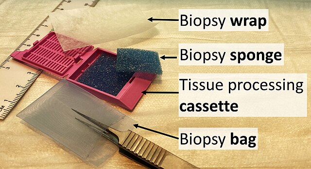
Items used for submitting specimens: (Biopsy) wrap, (biopsy) sponge, (tissue processing) cassette and (biopsy) bag.
There is also the option to make a "touch prep", wherein a glass slide is simply pressed against the tissue and then exposed to a fixative solution. The glass slide can then be stained and examined. This is feasible for an initial evaluation of suspected lymphomas.
Orientation (lowest magnification): In this case oriented by the skin surface (green). A lesion is seen (red) and its demarcation can be discerned (diffuse in this case)