The cerebral cortex, also known as the cerebral mantle, is the outer layer of neural tissue of the cerebrum of the brain in humans and other mammals. It is the largest site of neural integration in the central nervous system, and plays a key role in attention, perception, awareness, thought, memory, language, and consciousness. The cerebral cortex is the part of the brain responsible for cognition.
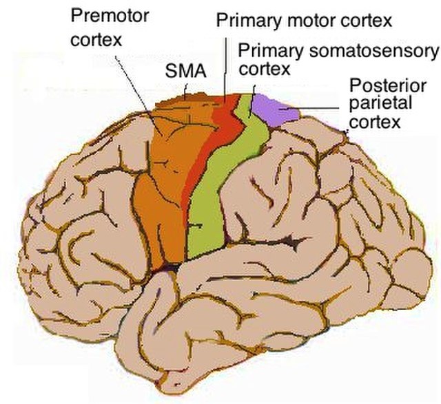
Lateral view of cerebrum showing several cortices
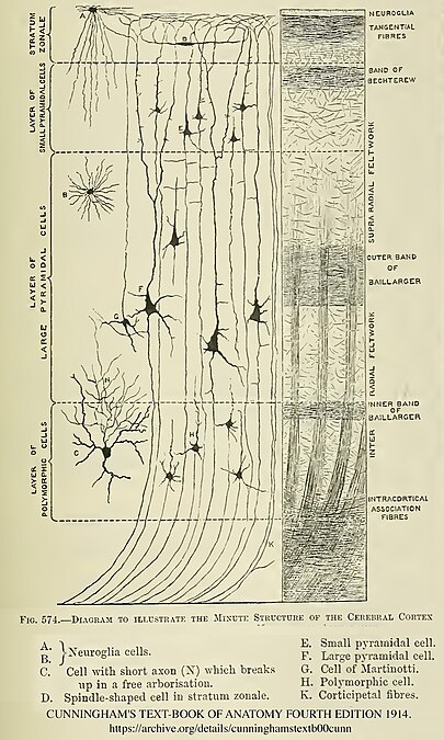
Diagram of layers pattern. Cells grouped on left, axonal layers on right.
Micrograph showing the visual cortex (predominantly pink). Subcortical white matter (predominantly blue) is seen at the bottom of the image. HE-LFB stain.
Cortical blood supply
The brain is an organ that serves as the center of the nervous system in all vertebrate and most invertebrate animals. In vertebrates, a small part of the brain called the hypothalamus is the neural control center for all endocrine systems. The brain is the largest cluster of neurons in the body and is typically located in the head, usually near organs for special senses such as vision, hearing and olfaction.
It is the most energy-consuming organ of the body, and the most specialized, responsible for endocrine regulation, sensory perception, motor control, and the development of intelligence.
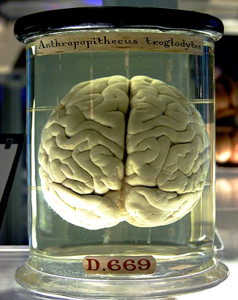
The brain of a chimpanzee
Cross section of the olfactory bulb of a rat, stained in two different ways at the same time: one stain shows neuron cell bodies, the other shows receptors for the neurotransmitter GABA.
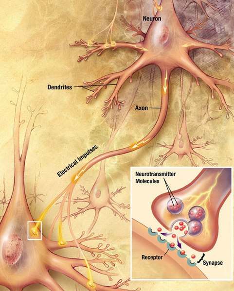
Neurons generate electrical signals that travel along their axons. When a pulse of electricity reaches a junction called a synapse, it causes a neurotransmitter chemical to be released, which binds to receptors on other cells and thereby alters their electrical activity.
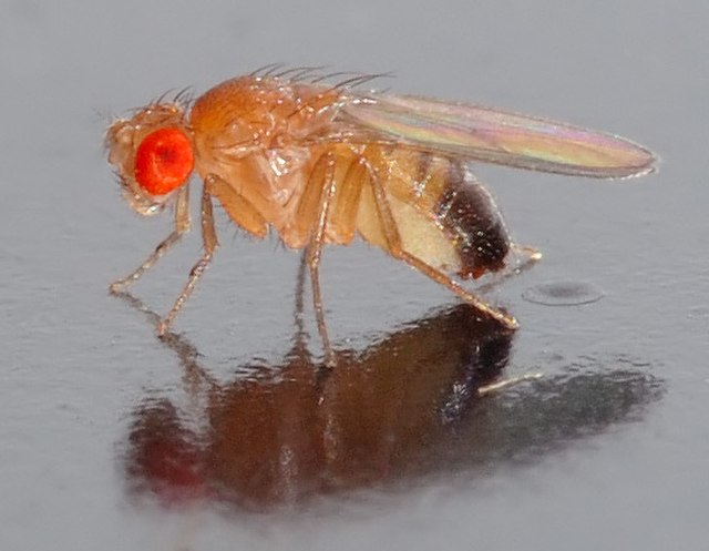
Fruit flies (Drosophila) have been extensively studied to gain insight into the role of genes in brain development.

