Dendritic cell
Videos
Page
A dendritic cell (DC) is an antigen-presenting cell of the mammalian immune system. A DC's main function is to process antigen material and present it on the cell surface to the T cells of the immune system. They act as messengers between the innate and adaptive immune systems.

Dendritic cells in skin

Artistic rendering of the surface of a human dendritic cell illustrating sheet-like processes that fold back onto the membrane surface. Some researchers believe that these sheets, when exposed to HIV, entrap viruses in the vicinity and focus them to contact zones with T cells targeted for infection. These studies were carried out using ion abrasion scanning electron microscopy, a new[when?] technology the NIH has been developing and applying for 3D cellular imaging.
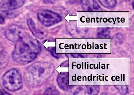
Histologic comparison of cell types in a germinal center, including follicular dendritic cells, H&E stain: - Centrocytes are small to medium size with angulated, elongated, cleaved, or twisted nuclei. - Centroblasts are larger cells containing vesicular nuclei with one to three basophilic nucleoli apposing the nuclear membrane. - Follicular dendritic cells have round nuclei, centrally located nucleoli, bland and dispersed chromatin, and flattening of adjacent nuclear membrane.

A dendritic cell
Immune system
Videos
Page
The immune system is a network of biological systems that protects an organism from diseases. It detects and responds to a wide variety of pathogens, from viruses to parasitic worms, as well as cancer cells and objects such as wood splinters, distinguishing them from the organism's own healthy tissue. Many species have two major subsystems of the immune system. The innate immune system provides a preconfigured response to broad groups of situations and stimuli. The adaptive immune system provides a tailored response to each stimulus by learning to recognize molecules it has previously encountered. Both use molecules and cells to perform their functions.

A scanning electron microscope image of a single neutrophil (yellow/right), engulfing anthrax bacteria (orange/left) – scale bar is 5 µm (false color)
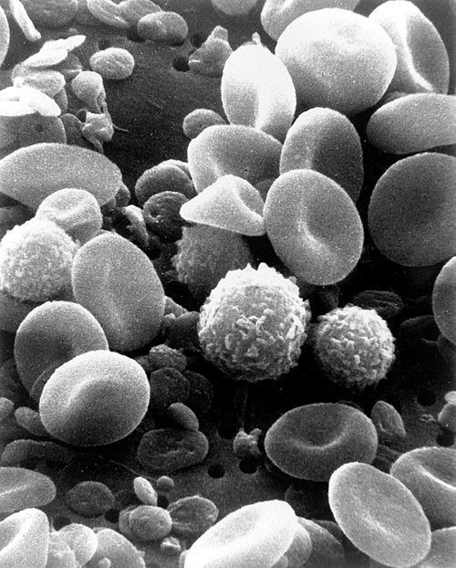
A scanning electron microscope image of normal circulating human blood. One can see red blood cells, several knobby white blood cells including lymphocytes, a monocyte, a neutrophil, and many small disc-shaped platelets.

An antibody is made up of two heavy chains and two light chains. The unique variable region allows an antibody to recognize its matching antigen.

Four neutrophils in a Giemsa-stained blood film