Silver staining
Videos
Page
In pathology, silver staining is the use of silver to selectively alter the appearance of a target in microscopy of histological sections; in temperature gradient gel electrophoresis; and in polyacrylamide gels.
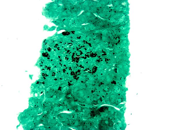
A silver stain (GMS) demonstrating the fungus Histoplasma (black round balls) in a liver biopsy.

RBC membrane proteins separated by SDS-PAGE and silver-stained.
Histology
Videos
Page
Histology,
also known as microscopic anatomy or microanatomy, is the branch of biology that studies the microscopic anatomy of biological tissues. Histology is the microscopic counterpart to gross anatomy, which looks at larger structures visible without a microscope. Although one may divide microscopic anatomy into organology, the study of organs, histology, the study of tissues, and cytology, the study of cells, modern usage places all of these topics under the field of histology. In medicine, histopathology is the branch of histology that includes the microscopic identification and study of diseased tissue. In the field of paleontology, the term paleohistology refers to the histology of fossil organisms.

Histologic specimen being placed on the stage of an optical microscope.

Human lung tissue stained with hematoxylin and eosin as seen under a microscope.

Histologic section of a plant stem (Alliaria petiolata).

Histologic section of a fossilized invertebrate. Ordovician bryozoan.