White blood cell
Videos
Page
White blood cells, also called immune cells or immunocytes, are cells of the immune system that are involved in protecting the body against both infectious disease and foreign invaders. White blood cells include three main subtypes: granulocytes, lymphocytes and monocytes.
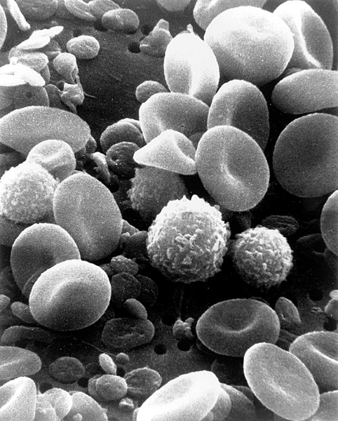
A scanning electron microscope image of normal circulating human blood. In addition to the irregularly shaped leukocytes, both red blood cells and many small disc-shaped platelets are visible.
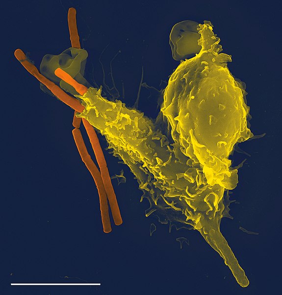
Neutrophil engulfing anthrax bacteria
Immune system
Videos
Page
The immune system is a network of biological systems that protects an organism from diseases. It detects and responds to a wide variety of pathogens, from viruses to parasitic worms, as well as cancer cells and objects such as wood splinters, distinguishing them from the organism's own healthy tissue. Many species have two major subsystems of the immune system. The innate immune system provides a preconfigured response to broad groups of situations and stimuli. The adaptive immune system provides a tailored response to each stimulus by learning to recognize molecules it has previously encountered. Both use molecules and cells to perform their functions.
A scanning electron microscope image of a single neutrophil (yellow/right), engulfing anthrax bacteria (orange/left) – scale bar is 5 µm (false color)
A scanning electron microscope image of normal circulating human blood. One can see red blood cells, several knobby white blood cells including lymphocytes, a monocyte, a neutrophil, and many small disc-shaped platelets.

An antibody is made up of two heavy chains and two light chains. The unique variable region allows an antibody to recognize its matching antigen.

Four neutrophils in a Giemsa-stained blood film