Apoptosis
Videos
Apoptosis is a form of programmed cell death that occurs in multicellular organisms and in some eukaryotic, single-celled microorganisms such as yeast. Biochemical events lead to characteristic cellul
An etoposide-treated DU145 prostate cancer cell exploding into a cascade of apoptotic bodies. The sub images were extracted from a 61-hour time-lapse microscopy video, created using quantitative phase-contrast microscopy. The optical thickness is color-coded. With increasing thickness, color changes from gray to yellow, red, purple and finally black. See the video at The Cell: An Image Library


John Sulston won the Nobel Prize in Medicine in 2002, for his pioneering research on apoptosis.

A section of mouse liver showing several apoptotic cells, indicated by arrows
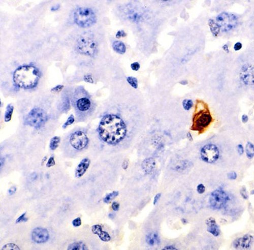
A section of mouse liver stained to show cells undergoing apoptosis (orange)
Immortalised cell line
Videos
An immortalised cell line is a population of cells from a multicellular organism that would normally not proliferate indefinitely but, due to mutation, have evaded normal cellular senescence and inste
Apoptosis
…be seen in the development of a lung cancer called NCI-H460. The X-linked inhibitor of apoptosis protein (XIAP) is overexpressed in cells of the H460 cell line. XIAPs bind to the processed form of caspase-9 and suppress the activity of apoptotic activator cytochrome c; therefore, overexpression leads to a…
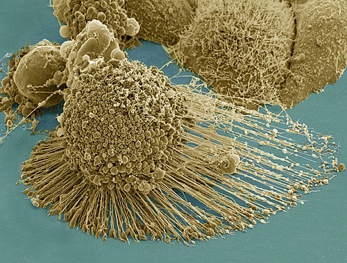
Scanning electron micrograph of an apoptotic HeLa cell. Zeiss Merlin HR-SEM.
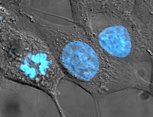
HeLa cells, an example of an immortalised cell line. DIC image, DNA stained with Hoechst 33258.