Atrium (heart)
Videos
The atrium is one of the two upper chambers in the heart that receives blood from the circulatory system. The blood in the atria is pumped into the heart ventricles through the atrioventricular mitral
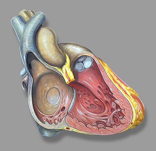
Right heart anatomy, right ventricle seen on right of illustration
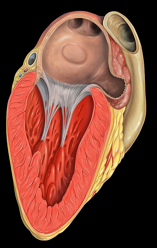
Left atrial appendage shown at upper right

CT scan of the chest showing a thrombus in the left atrial appendage (left: axial plane, right: coronal plane)
Heart
Videos
The heart is a muscular organ found in humans and other animals. This organ pumps blood through the blood vessels. The heart and blood vessels together make up the circulatory system. The pumped blood
Atrium (heart)
…The atrium (Latin: ātrium, lit. 'entry hall'; pl.: atria) is one of the two upper chambers in the heart that receives blood from the circulatory system. The blood in the atria is pumped into the heart ventricles through the atrioventricular…

Heart being dissected showing right and left ventricles, from above

With the atria and major vessels removed, all four valves are clearly visible.

Frontal section showing papillary muscles attached to the tricuspid valve on the right and to the mitral valve on the left via chordae tendineae.

Layers of the heart wall, including visceral and parietal pericardium