Autofluorescence is the natural emission of light by biological structures such as mitochondria and lysosomes when they have absorbed light, and is used to distinguish the light originating from artificially added fluorescent markers (fluorophores).
Micrograph of paper autofluorescing under ultraviolet illumination. The individual fibres in this sample are around 10 μm in diameter.
Autofluorescence super resolution microscopy/optical nanoscopy image of cellular structures that are invisible with confocal light microscopy
Autofluorescence in banana skin under different light conditions.
A fluorescence microscope is an optical microscope that uses fluorescence instead of, or in addition to, scattering, reflection, and attenuation or absorption, to study the properties of organic or inorganic substances. "Fluorescence microscope" refers to any microscope that uses fluorescence to generate an image, whether it is a simple set up like an epifluorescence microscope or a more complicated design such as a confocal microscope, which uses optical sectioning to get better resolution of the fluorescence image.
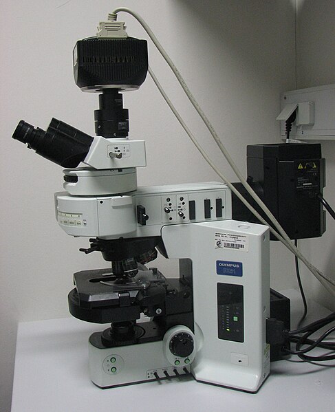
An upright fluorescence microscope (Olympus BX61) with the fluorescence filter cube turret above the objective lenses, coupled with a digital camera.
A sample of herring sperm stained with SYBR green in a cuvette illuminated by blue light in an epifluorescence microscope. The SYBR green in the sample binds to the herring sperm DNA and, once bound, fluoresces giving off green light when illuminated by blue light.
Epifluorescent imaging of the three components in a dividing human cancer cell. DNA is stained blue, a protein called INCENP is green, and the microtubules are red. Each fluorophore is imaged separately using a different combination of excitation and emission filters, and the images are captured sequentially using a digital CCD camera, then overlaid to give a complete image.
Endothelial cells under the microscope. Nuclei are stained blue with DAPI, microtubules are marked green by an antibody bound to FITC and actin filaments are labeled red with phalloidin bound to TRITC. Bovine pulmonary artery endothelial (BPAE) cells