Autophagy
Videos
Autophagy is the natural, conserved degradation of a biological cell that removes unnecessary or dysfunctional components through a lysosome-dependent regulated mechanism. It allows the orderly degrad
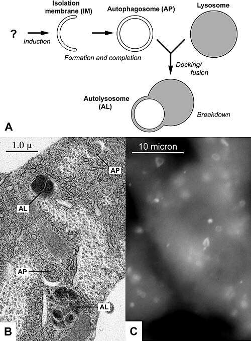
A Diagram of the process of autophagy, which produces the structures autophagosomes (AP), and autolysosomes (AL) B Electron micrograph of autophagic structures AP and AL in the fat body of a fruit fly larva C Fluorescently labeled autophagosomes AP in liver cells of starved mice
Lysosome
Videos
A lysosome is a membrane-bound organelle that is found in all animal cells, and rarely in plant cells. There are normally hundreds of lysosomes in the cytosol, where they function as the cell's degrad
Autophagy
…kýtos, meaning "hollow") is the natural, conserved degradation of a biological cell that removes unnecessary or dysfunctional components through a lysosome-dependent regulated mechanism. It allows the orderly degradation and recycling of cellular components. Although initially characterized as a primordial…

TEM views of various vesicular compartments. Lysosomes are denoted by "Ly". They are dyed dark due to their acidity; in the center of the top image, a Golgi Apparatus can be seen, distal from the cell membrane relative to the lysosome.

Cellular material is delivered to lysosomes in four different ways: (A) Macroautophagy, (B) Endosomal degradation, (C) Microautophagy and (D) Chaperone-mediated autophagy (CMA).

Christian de Duve, age 95, presenting his ideas on the origin of the eukaryotic cell (October 2012)