Cell migration is a central process in the development and maintenance of multicellular organisms. Tissue formation during embryonic development, wound healing and immune responses all require the orchestrated movement of cells in particular directions to specific locations. Cells often migrate in response to specific external signals, including chemical signals and mechanical signals. Errors during this process have serious consequences, including intellectual disability, vascular disease, tumor formation and metastasis. An understanding of the mechanism by which cells migrate may lead to the development of novel therapeutic strategies for controlling, for example, invasive tumour cells.

(A) Dynamic microtubules are necessary for tail retraction and are distributed at the rear end in a migrating cell. Green, highly dynamic microtubules; yellow, moderately dynamic microtubules and red, stable microtubules. (B) Stable microtubules act as struts and prevent tail retraction and thereby inhibit cell migration.
Schematic representation of the collective biomechanical and molecular mechanism of cell motion
Rearward membrane flow (red arrows) and vesicle trafficking from back to front (blue arrows) drive adhesion-independent migration.
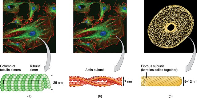
The cytoskeleton is a complex, dynamic network of interlinking protein filaments present in the cytoplasm of all cells, including those of bacteria and archaea. In eukaryotes, it extends from the cell nucleus to the cell membrane and is composed of similar proteins in the various organisms. It is composed of three main components: microfilaments, intermediate filaments, and microtubules, and these are all capable of rapid growth or disassembly depending on the cell's requirements.
The cytoskeleton consists of (a) microtubules, (b) microfilaments, and (c) intermediate filaments.
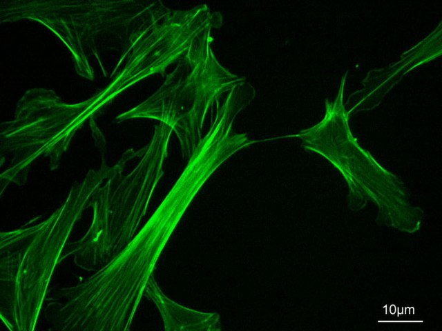
Actin cytoskeleton of mouse embryo fibroblasts, stained with phalloidin
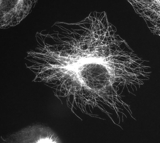
Microtubules in a gel-fixated cell
Cross section diagram through the cilium, showing the “9 + 2” arrangement of microtubules


