Cell nucleus
Videos
The cell nucleus is a membrane-bound organelle found in eukaryotic cells. Eukaryotic cells usually have a single nucleus, but a few cell types, such as mammalian red blood cells, have no nuclei, and a
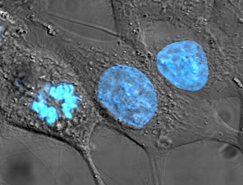
HeLa cells stained for nuclear DNA with the blue fluorescent Hoechst dye. The central and rightmost cells are in interphase, thus their entire nuclei are labeled. On the left, a cell is going through mitosis and its DNA has condensed.

An image of a newt lung cell stained with fluorescent dyes during metaphase. The mitotic spindle can be seen, stained green, attached to the two sets of chromosomes, stained light blue. All chromosomes but one are already at the metaphase plate.

Oldest known depiction of cells and their nuclei by Antonie van Leeuwenhoek, 1719

Drawing of a Chironomus salivary gland cell published by Walther Flemming in 1882. The nucleus contains polytene chromosomes.
Organelle
Videos
An organelle is a specialized subunit, within a biological cell, that has a specific function. The name organelle comes from the idea that these structures are parts of cells, as organs are to the bod
Cell nucleus
…The cell nucleus (from Latin nucleus or nuculeus 'kernel, seed'; pl.: nuclei) is a membrane-bound organelle found in eukaryotic cells. Eukaryotic cells usually have a single nucleus, but a few cell types, such as mammalian red blood cells, have no nuclei, and…

(A) Electron micrograph of Halothiobacillus neapolitanus cells, arrows highlight carboxysomes. (B) Image of intact carboxysomes isolated from H. neapolitanus. Scale bars are 100 nm.

Structure of Candidatus Brocadia anammoxidans, showing an anammoxosome and intracytoplasmic membrane

Diagram showing the organelles of a generalised animal cell