A cnidocyte is an explosive cell containing one large secretory organelle called a cnidocyst that can deliver a sting to other organisms. The presence of this cell defines the phylum Cnidaria. Cnidae are used to capture prey and as a defense against predators. A cnidocyte fires a structure that contains a toxin within the cnidocyst; this is responsible for the stings delivered by a cnidarian. Cnidocytes are single-use cells that need to be continuously replaced.
Nomarski micrograph of a ruthenium red-stained nematocyst from Aiptasia pallida, the pale anemone. The red dye stains the polyanionic venom proteins found inside the capsule of this partially-discharged nematocyst.
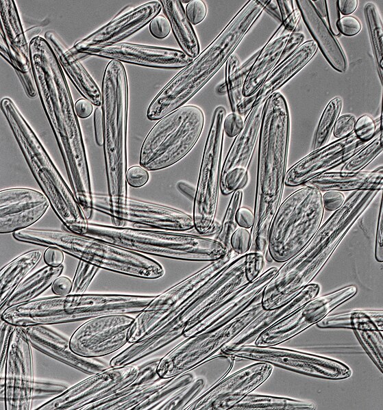
Nematocysts from Chironex fleckeri (400x magnification)
In cell biology, an organelle is a specialized subunit, usually within a cell, that has a specific function. The name organelle comes from the idea that these structures are parts of cells, as organs are to the body, hence organelle, the suffix -elle being a diminutive. Organelles are either separately enclosed within their own lipid bilayers or are spatially distinct functional units without a surrounding lipid bilayer. Although most organelles are functional units within cells, some function units that extend outside of cells are often termed organelles, such as cilia, the flagellum and archaellum, and the trichocyst.
(A) Electron micrograph of Halothiobacillus neapolitanus cells, arrows highlight carboxysomes. (B) Image of intact carboxysomes isolated from H. neapolitanus. Scale bars are 100 nm.
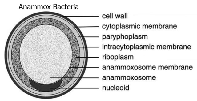
Structure of Candidatus Brocadia anammoxidans, showing an anammoxosome and intracytoplasmic membrane