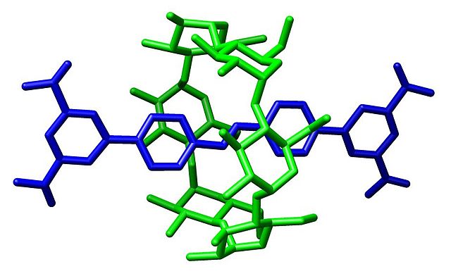
A cystine knot is a protein structural motif containing three disulfide bridges. The sections of polypeptide that occur between two of them form a loop through which a third disulfide bond passes, forming a rotaxane substructure. The cystine knot motif stabilizes protein structure and is conserved in proteins across various species. There are three types of cystine knot, which differ in the topology of the disulfide bonds:The growth factor cystine knot (GFCK)
inhibitor cystine knot (ICK) common in spider and snail toxins
Cyclic Cystine Knot, or cyclotide
Structure of human chorionic gonadotropin.
A rotaxane is a mechanically interlocked molecular architecture consisting of a dumbbell-shaped molecule which is threaded through a macrocycle. The two components of a rotaxane are kinetically trapped since the ends of the dumbbell are larger than the internal diameter of the ring and prevent dissociation (unthreading) of the components since this would require significant distortion of the covalent bonds.
Graphical representation of a rotaxane
(a) A rotaxane is formed from an open ring (R1) with a flexible hinge and a dumbbell-shaped DNA origami structure (D1). The hinge of the ring consists of a series of strand crossovers into which additional thymines are inserted to provide higher flexibility. Ring and axis subunits are first connected and positioned with respect to each other using 18 nucleotide long, complementary sticky ends 33 nm away from the center of the axis (blue regions). The ring is then closed around the dumbbell axis using closing strands (red), followed by the addition of release strands that separate dumbbell from ring via toehold-mediated strand displacement. (b) 3D models and corresponding averaged
Structure of a rotaxane with an α-cyclodextrin macrocycle.


