A dendritic spine is a small membranous protrusion from a neuron's dendrite that typically receives input from a single axon at the synapse. Dendritic spines serve as a storage site for synaptic strength and help transmit electrical signals to the neuron's cell body. Most spines have a bulbous head, and a thin neck that connects the head of the spine to the shaft of the dendrite. The dendrites of a single neuron can contain hundreds to thousands of spines. In addition to spines providing an anatomical substrate for memory storage and synaptic transmission, they may also serve to increase the number of possible contacts between neurons. It has also been suggested that changes in the activity of neurons have a positive effect on spine morphology.
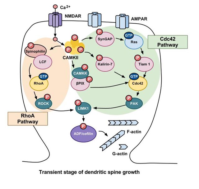
Calcium influx through NMDA receptors activates CAMKII. CAMKII then regulates several other signaling cascades that modulate the activity of the actin-binding proteins cofilin and profilin. These cascades can be divided into two primary pathways, the RhoA and Cdc42 pathways, which are mediated primarily by these members of the Rho family of GTPases. In the transient stage, the signaling cascade caused by synaptic activity results in LIMK1 phosphorylating ADF/cofilin via both the RhoA and Cdc42 pathways, which in turn inhibits the depolymerization of F-actin and increases the volume of the dendritic spine drastically while also inducing LTP.

In contrast, the sustained stage is focused more on activating the RhoA pathway, which ultimately results in a higher concentration of profilin, which prevents additional polymerization of actin and decreases the size of the dendritic spine from the transient stage, though still allows it to remain at an elevated level compared to an unpotentiated spine.
Experience-dependent spine formation and elimination
An excitatory synapse is a synapse in which an action potential in a presynaptic neuron increases the probability of an action potential occurring in a postsynaptic cell. Neurons form networks through which nerve impulses travels, each neuron often making numerous connections with other cells of neurons. These electrical signals may be excitatory or inhibitory, and, if the total of excitatory influences exceeds that of the inhibitory influences, the neuron will generate a new action potential at its axon hillock, thus transmitting the information to yet another cell.
Histological brain sample of the Substantia Nigra in Parkinson's disease, showing the presence of Lewy bodies and other signs of neurodegeneration.


