Epiphyseal plate
Videos
The epiphyseal plate, epiphysial plate, physis, or growth plate is a hyaline cartilage plate in the metaphysis at each end of a long bone. It is the part of a long bone where new bone growth takes pla
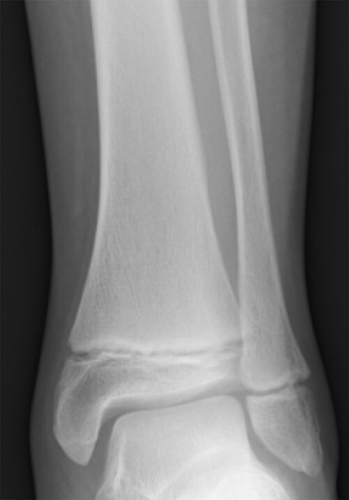
Radiogram of distal tibia (left) and fibula (right) showing two epiphyseal plates

Light micrograph of an undecalcified epiphyseal plate that is displaying the hypertrophic zone with its typical chondrocytes, matrix and three zones: maturation (top), degenerative (middle) and provisional calcification (bottom).

Masson Goldner trichrome stain of rabbit epiphysial plate
Hyaline cartilage
Videos
Hyaline cartilage is the glass-like and translucent cartilage found on many joint surfaces. It is also most commonly found in the ribs, nose, larynx, and trachea. Hyaline cartilage is pearl-gray in co
Epiphyseal plate
…The epiphyseal plate, epiphysial plate, physis, or growth plate is a hyaline cartilage plate in the metaphysis at each end of a long bone. It is the part of a long bone where new bone growth takes place; that is, the whole bone is alive…

Histology of articular cartilage zones.

Elbow joint. Deep dissection. Anterior view.