HeLa is an immortalized cell line used in scientific research. It is the oldest human cell line and one of the most commonly used. HeLa cells are durable and prolific, allowing for extensive applications in scientific study. The line is derived from cervical cancer cells taken on 8 February 1951, from Henrietta Lacks, a 31-year-old African American mother of five, after whom the line is named. Lacks died of cancer on 4 October 1951.
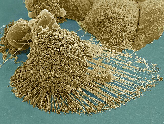
Scanning electron micrograph of an apoptotic HeLa cell. Zeiss Merlin HR-SEM.
Multiphoton fluorescence image of cultured HeLa cells with a fluorescent protein targeted to the Golgi apparatus (orange), microtubules (green) and counterstained for DNA (cyan). Nikon RTS2000MP custom laser scanning microscope.
Immunofluorescence image of HeLa cells grown in tissue culture and stained with antibody to actin in green, vimentin in red and DNA in blue
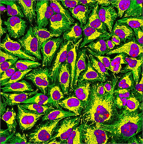
Immunofluorescence of HeLa cells showing microtubules in green, mitochondria in yellow, nucleoli in red and nuclear DNA in purple
An immortalised cell line is a population of cells from a multicellular organism which would normally not proliferate indefinitely but, due to mutation, have evaded normal cellular senescence and instead can keep undergoing division. The cells can therefore be grown for prolonged periods in vitro. The mutations required for immortality can occur naturally or be intentionally induced for experimental purposes. Immortal cell lines are a very important tool for research into the biochemistry and cell biology of multicellular organisms. Immortalised cell lines have also found uses in biotechnology.
Scanning electron micrograph of an apoptotic HeLa cell. Zeiss Merlin HR-SEM.
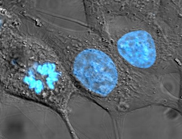
HeLa cells, an example of an immortalised cell line. DIC image, DNA stained with Hoechst 33258.