Histopathology is the microscopic examination of tissue in order to study the manifestations of disease. Specifically, in clinical medicine, histopathology refers to the examination of a biopsy or surgical specimen by a pathologist, after the specimen has been processed and histological sections have been placed onto glass slides. In contrast, cytopathology examines free cells or tissue micro-fragments.
Micrograph showing contraction band necrosis, a histopathologic finding of myocardial infarction (heart attack).
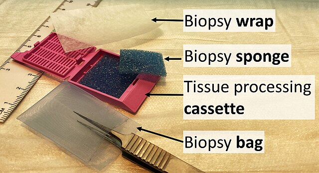
Items used for submitting specimens: (Biopsy) wrap, (biopsy) sponge, (tissue processing) cassette and (biopsy) bag.
There is also the option to make a "touch prep", wherein a glass slide is simply pressed against the tissue and then exposed to a fixative solution. The glass slide can then be stained and examined. This is feasible for an initial evaluation of suspected lymphomas.
Orientation (lowest magnification): In this case oriented by the skin surface (green). A lesion is seen (red) and its demarcation can be discerned (diffuse in this case)
The optical microscope, also referred to as a light microscope, is a type of microscope that commonly uses visible light and a system of lenses to generate magnified images of small objects. Optical microscopes are the oldest design of microscope and were possibly invented in their present compound form in the 17th century. Basic optical microscopes can be very simple, although many complex designs aim to improve resolution and sample contrast.
Scientist using an optical microscope in a laboratory
A miniature USB microscope
The oldest published image known to have been made with a microscope: bees by Francesco Stelluti, 1630
Basic optical transmission microscope elements (1990s)