Tooth development or odontogenesis is the complex process by which teeth form from embryonic cells, grow, and erupt into the mouth. For human teeth to have a healthy oral environment, all parts of the tooth must develop during appropriate stages of fetal development. Primary (baby) teeth start to form between the sixth and eighth week of prenatal development, and permanent teeth begin to form in the twentieth week. If teeth do not start to develop at or near these times, they will not develop at all, resulting in hypodontia or anodontia.
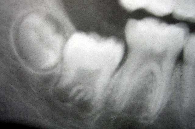
Radiograph of lower right (from left to right) third, second, and first molars in different stages of development
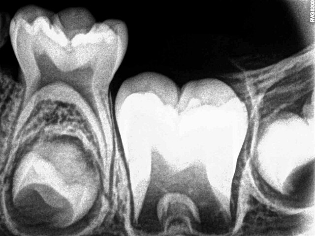
X-ray of teeth of a boy aged 5 years showing left lower primary molar and developing crowns of left lower permanent premolar (below primary molar) and permanent molars
Histologic slide showing a tooth bud. A: enamel organ B: dental papilla C: dental follicle
Histology of important stages of tooth development
A tooth is a hard, calcified structure found in the jaws of many vertebrates and used to break down food. Some animals, particularly carnivores and omnivores, also use teeth to help with capturing or wounding prey, tearing food, for defensive purposes, to intimidate other animals often including their own, or to carry prey or their young. The roots of teeth are covered by gums. Teeth are not made of bone, but rather of multiple tissues of varying density and hardness that originate from the outermost embryonic germ layer, the ectoderm.
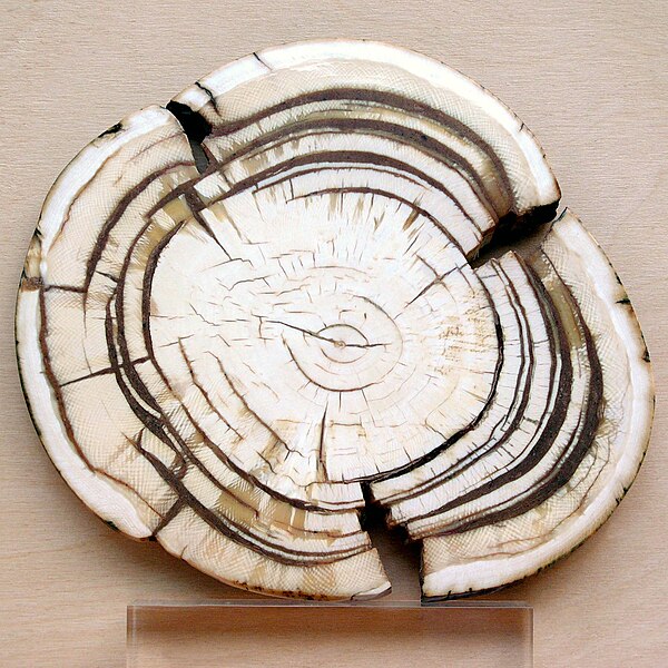
Section through the ivory tusk of a mammoth
Buccal view of top incisor from Rattus rattus. Top incisor outlined in yellow. Molars circled in blue.
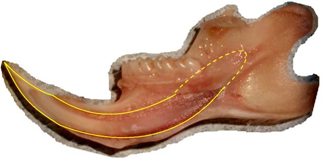
Buccal view of the lower incisor from the right dentary of a Rattus rattus
Lingual view of the lower incisor from the right dentary of a Rattus rattus


