Immune system
Videos
Photos
The immune system is a network of biological systems that protects an organism from diseases. It detects and responds to a wide variety of pathogens, from viruses to parasitic worms, as well as cancer cells and objects such as wood splinters, distinguishing them from the organism's own healthy tissue. Many species have two major subsystems of the immune system. The innate immune system provides a preconfigured response to broad groups of situations and stimuli. The adaptive immune system provides a tailored response to each stimulus by learning to recognize molecules it has previously encountered. Both use molecules and cells to perform their functions.

A scanning electron microscope image of a single neutrophil (yellow/right), engulfing anthrax bacteria (orange/left) – scale bar is 5 µm (false color)
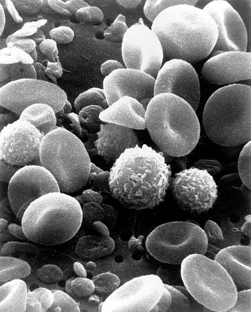
A scanning electron microscope image of normal circulating human blood. One can see red blood cells, several knobby white blood cells including lymphocytes, a monocyte, a neutrophil, and many small disc-shaped platelets.

An antibody is made up of two heavy chains and two light chains. The unique variable region allows an antibody to recognize its matching antigen.

Four neutrophils in a Giemsa-stained blood film
Parasitic worm
Videos
Photos
Parasitic worms, also known as helminths, are large macroparasites; adults can generally be seen with the naked eye. Many are intestinal worms that are soil-transmitted and infect the gastrointestinal tract. Other parasitic worms such as schistosomes reside in blood vessels.

Hookworms attached to the intestinal mucosa

Two pinworms

Processed helminth eggs samples from a dry toilet in Kenya

Analysing for helminth eggs in samples of feces from a dry toilet in Kenya