Immunofluorescence (IF) is a light microscopy-based technique that allows detection and localization of a wide variety of target biomolecules within a cell or tissue at a quantitative level. The technique utilizes the binding specificity of antibodies and antigens. The specific region an antibody recognizes on an antigen is called an epitope. Several antibodies can recognize the same epitope but differ in their binding affinity. The antibody with the higher affinity for a specific epitope will surpass antibodies with a lower affinity for the same epitope.
Vasculature of porcine skin under fluorescence (Smooth muscle actin with AlexaFluor 488). Green = smooth muscle actin (SMA) with Alexa 488 fluorophore. Blue = DAPI counterstain. Red = auto-fluorescence.
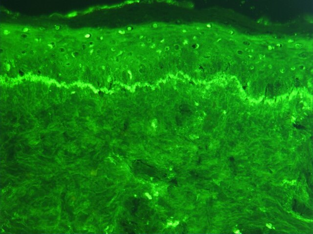
Photomicrograph of a histological section of human skin prepared for direct immunofluorescence using an anti-IgG antibody. The skin is from a patient with systemic lupus erythematosus and shows IgG deposit at two different places: The first is a band-like deposit along the epidermal basement membrane ("lupus band test" is positive). The second is within the nuclei of the epidermal cells (anti-nuclear antibodies).
Basic concept of the LSAB-method: Utilizes a Streptavidin–enzyme conjugate for the identification of the biotinylated secondary antibody which is bound to the primary antibody. This approach is applicable when the Avidin–Biotin complex in the ABC method becomes too large.
Photomicrograph of a histological section of human skin prepared for direct immunofluorescence using an anti-IgA antibody. The skin is from a patient with Henoch–Schönlein purpura: IgA deposits are found in the walls of small superficial capillaries (yellow arrows). The pale wavy green area on top is the epidermis, the bottom fibrous area is the dermis.
The optical microscope, also referred to as a light microscope, is a type of microscope that commonly uses visible light and a system of lenses to generate magnified images of small objects. Optical microscopes are the oldest design of microscope and were possibly invented in their present compound form in the 17th century. Basic optical microscopes can be very simple, although many complex designs aim to improve resolution and sample contrast.
Scientist using an optical microscope in a laboratory
A miniature USB microscope
The oldest published image known to have been made with a microscope: bees by Francesco Stelluti, 1630
Basic optical transmission microscope elements (1990s)


