Immunohistochemistry (IHC) is a form of immunostaining. It involves the process of selectively identifying antigens (proteins) in cells and tissue, by exploiting the principle of antibodies binding specifically to antigens in biological tissues. Albert Hewett Coons, Ernest Berliner, Norman Jones and Hugh J Creech was the first to develop immunofluorescence in 1941. This led to the later development of immunohistochemistry.
Main staining patterns on chromogenic immunohistochemistry.
Immunofluorescence of human skin using an anti-IgA antibody. The skin is from a patient with Henoch–Schönlein purpura: IgA deposits are found in the walls of small superficial capillaries (yellow arrows). The pale wavy green area on top is the epidermis, the bottom fibrous area is the dermis.
Chromogenic immunohistochemistry: The cell is exposed to a primary antibody (red) that binds to a specific antigen (purple square). The primary antibody binds a secondary (green) antibody that is chemically coupled to an enzyme (blue). The enzyme changes the color of the substrate to a more pigmented one (brown star).
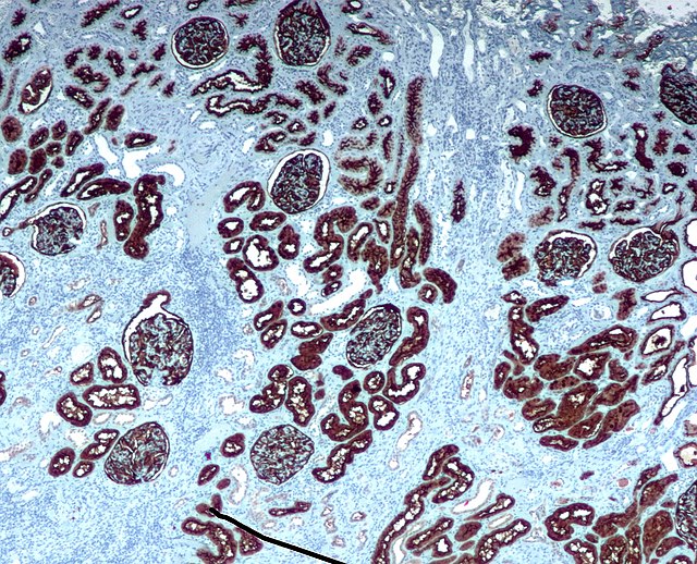
Chromogenic immunohistochemistry of a normal kidney targeting the protein CD10.
In biochemistry, immunostaining is any use of an antibody-based method to detect a specific protein in a sample. The term "immunostaining" was originally used to refer to the immunohistochemical staining of tissue sections, as first described by Albert Coons in 1941. However, immunostaining now encompasses a broad range of techniques used in histology, cell biology, and molecular biology that use antibody-based staining methods.
Micrograph of a GFAP immunostained section of a brain tumour.