Large intestine
Videos
The large intestine, also known as the large bowel, is the last part of the gastrointestinal tract and of the digestive system in tetrapods. Water and salts are absorbed here and the remaining waste m

Colonic crypts (intestinal glands) within four tissue sections. The cells have been stained to show a brown-orange color if the cells produce the mitochondrial protein cytochrome c oxidase subunit I (CCOI), and the nuclei of the cells (located at the outer edges of the cells lining the walls of the crypts) are stained blue-gray with haematoxylin. Panels A, B were cut across the long axes of the crypts and panels C, D were cut parallel to the long axes of the crypts. In panel A the bar shows 100 μm and allows an estimate of the frequency of crypts in the colonic epithelium. Panel B includes three crypts in cross-section, each with one segment deficient for CCOI expression and at least one crypt, on the right side, undergoing fission into two crypts. Panel C shows, on the left side, a crypt fissioning into two crypts. Panel D shows typical small clusters of two and three CCOI deficient crypts (the bar shows 50 μm). The images were made from original photomicrographs, but panels A, B and D were also included in an article

Micrograph of normal large instestinal crypts.

Anatomy of normal large intestinal crypts

Histological section.
Cecum
Videos
The cecum is a pouch within the peritoneum that is considered to be the beginning of the large intestine. It is typically located on the right side of the body. The term stems from the Latin caecus, m
Large intestine
…medicine, the terms "large intestine" and "colon" are often used interchangeably, but most sources define the large intestine as the combination of the cecum, colon, rectum, and anal canal. Some other sources exclude the anal canal. The colon of the large intestine is the last part of the digestive system…

Cecum and ileum

Ileo-cecal valve
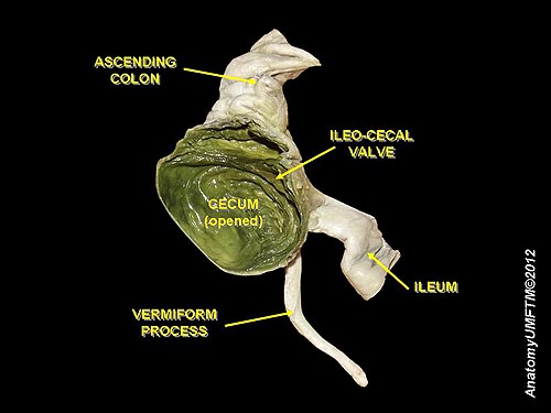
Cecum

Endoscopic image of cecum with arrow pointing to ileocecal valve in foreground