Leucine-rich repeat
Videos
A leucine-rich repeat is a protein structural motif that forms an α/β horseshoe fold. It is composed of repeating 20–30 amino acid stretches that are unusually rich in the hydrophobic amino acid leuci

a leucine-rich repeat variant with a novel repetitive protein structural motif

internalin h: crystal structure of fused n-terminal domains.

dimeric bovine tissue-extracted decorin, crystal form 2

the crystal structure of pgip (polygalacturonase inhibiting protein), a leucine rich repeat protein involved in plant defense
Alpha helix
Videos
An alpha helix is a sequence of amino acids in a protein that are twisted into a coil.
Leucine-rich repeat
…repeats commonly fold together to form a solenoid protein domain, termed leucine-rich repeat domain. Typically, each repeat unit has beta strand-turn-alpha helix structure, and the assembled domain, composed of many such repeats, has a horseshoe shape with an interior parallel beta sheet and an exterior array…

Contrast of helix end views between α (offset squarish) vs 310 (triangular)
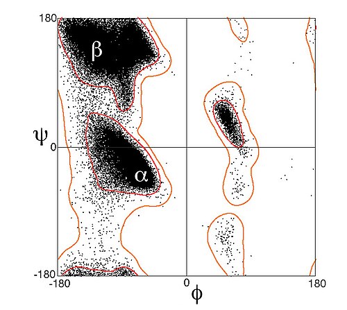
Ramachandran plot (φ, ψ plot), with data points for α-helical residues forming a dense diagonal cluster below and left of center, around the global energy minimum for backbone conformation.

An α-helix in ultrahigh-resolution electron density contours, with oxygen atoms in red, nitrogen atoms in blue, and hydrogen bonds as green dotted lines (PDB file 2NRL, 17–32). The N-terminus is at the top, here.

Leucine zipper coiled-coil helices & DNA-binding helices: transcription factor Max (PDB file 1HLO)