Locus coeruleus
Videos
The locus coeruleus, also spelled locus caeruleus or locus ceruleus, is a nucleus in the pons of the brainstem involved with physiological responses to stress and panic. It is a part of the reticular
Micrograph showing the locus coeruleus (upper-right of image) in an axial section of the pons. The fourth ventricle (quasi-triangular white area) is in the upper-left of the image. The midline is seen on the left. The large white area in the upper-left corner is where the cerebellum would be. HE-LFB stain.


Locus coeruleus highlighted in green.
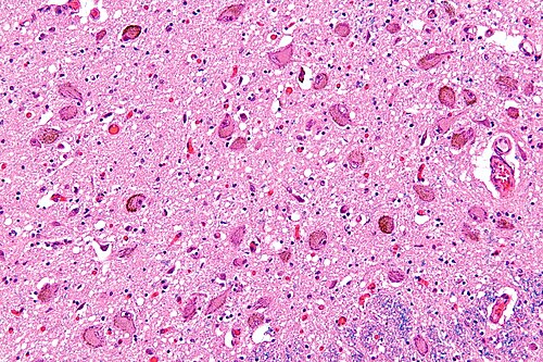
Micrograph showing the locus coeruleus. HE-LFB stain
Cingulate cortex
Videos
The cingulate cortex is a part of the brain situated in the medial aspect of the cerebral cortex. The cingulate cortex includes the entire cingulate gyrus, which lies immediately above the corpus call
Locus coeruleus
…to be activated by stimuli. As an important homeostatic control center of the body, the locus coeruleus receives afferents from the hypothalamus. The cingulate gyrus and the amygdala also innervate the LC, allowing emotional pain and stressors to trigger noradrenergic responses. The cerebellum and afferents from the…

Micrograph showing spindle neurons of the cingulate cortex. HE-LFB stain.

Medial sagittal section of the human brain.

Median sagittal section of the human brain.