A mitochondrion is an organelle found in the cells of most eukaryotes, such as animals, plants and fungi. Mitochondria have a double membrane structure and use aerobic respiration to generate adenosine triphosphate (ATP), which is used throughout the cell as a source of chemical energy. They were discovered by Albert von Kölliker in 1857 in the voluntary muscles of insects. The term mitochondrion was coined by Carl Benda in 1898. The mitochondrion is popularly nicknamed the "powerhouse of the cell", a phrase coined by Philip Siekevitz in a 1957 article of the same name.
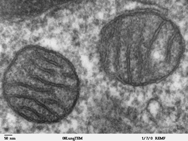
Two mitochondria from mammalian lung tissue displaying their matrix and membranes as shown by electron microscopy
Cross-sectional image of cristae in a rat liver mitochondrion to demonstrate the likely 3D structure and relationship to the inner membrane
Transmission electron micrograph of a chondrocyte, stained for calcium, showing its nucleus (N) and mitochondria (M)
Typical mitochondrial network (green) in two human cells (HeLa cells)
In cell biology, an organelle is a specialized subunit, usually within a cell, that has a specific function. The name organelle comes from the idea that these structures are parts of cells, as organs are to the body, hence organelle, the suffix -elle being a diminutive. Organelles are either separately enclosed within their own lipid bilayers or are spatially distinct functional units without a surrounding lipid bilayer. Although most organelles are functional units within cells, some function units that extend outside of cells are often termed organelles, such as cilia, the flagellum and archaellum, and the trichocyst.
(A) Electron micrograph of Halothiobacillus neapolitanus cells, arrows highlight carboxysomes. (B) Image of intact carboxysomes isolated from H. neapolitanus. Scale bars are 100 nm.
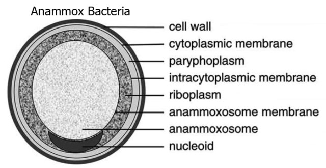
Structure of Candidatus Brocadia anammoxidans, showing an anammoxosome and intracytoplasmic membrane


