Occlusion, in a dental context, means simply the contact between teeth. More technically, it is the relationship between the maxillary (upper) and mandibular (lower) teeth when they approach each other, as occurs during chewing or at rest.
Anatomy of the temporomandibular joint - RCP = Here we see the condyle when teeth are in the retruded contact position, a reproducible position. ICP = Here we see the condyle position when teeth are in the intercuspal position. R = Mandibular opening with rotation of the condylar heads but without translation. T = Maximum opening of the mandible combined rotation and translation of condylar heads. (Institute of Dentistry, Aberdeen University)
Leeway space is the size differential between the primary posterior teeth (C,D,E) and the permanent teeth (canine, first and second pre-molar). Maxillary space of 1.5mm, mandibular 2.5mm can be seen. (Institute of Dentistry, Aberdeen University)
Molar relationship classification, observed when locating the mesial buccal cusp of the maxillary first molar and buccal groove of the mandibular first molar. (Institute of Dentistry, Aberdeen University)
Intercuspal Position -The relationship between the mandible and the maxilla when the teeth are maximally meshed. It is the most cranial position of the mandible (Institute of Dentistry Aberdeen University)
Tooth development or odontogenesis is the complex process by which teeth form from embryonic cells, grow, and erupt into the mouth. For human teeth to have a healthy oral environment, all parts of the tooth must develop during appropriate stages of fetal development. Primary (baby) teeth start to form between the sixth and eighth week of prenatal development, and permanent teeth begin to form in the twentieth week. If teeth do not start to develop at or near these times, they will not develop at all, resulting in hypodontia or anodontia.
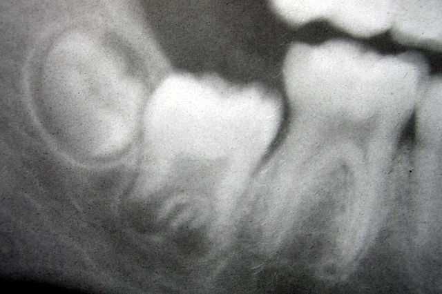
Radiograph of lower right (from left to right) third, second, and first molars in different stages of development
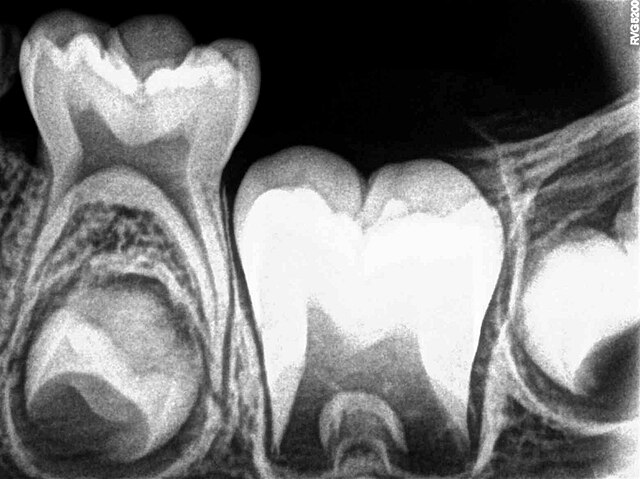
X-ray of teeth of a boy aged 5 years showing left lower primary molar and developing crowns of left lower permanent premolar (below primary molar) and permanent molars
Histologic slide showing a tooth bud. A: enamel organ B: dental papilla C: dental follicle
Histology of important stages of tooth development