Optogenetics
Videos
Optogenetics is a biological technique used to characterize and manipulate the activity of neurons or other cell types with light. This is achieved by expression of light-sensitive ion channels, pumps

Fig 1. Channelrhodopsin-2 (ChR2) induces temporally precise blue light-driven activity in rat prelimbic prefrontal cortical neurons. a) In vitro schematic (left) showing blue light delivery and whole-cell patch-clamp recording of light-evoked activity from a fluorescent CaMKllα::ChR2-EYFP expressing pyramidal neuron (right) in an acute brain slice. b) In vivo schematic (left) showing blue light (473 nm) delivery and single-unit recording. (bottom left) Coronal brain slice showing expression of CaMKllα::ChR2-EYFP in the prelimbic region. Light blue arrow shows tip of the optical fiber; black arrow shows tip of the recording electrode (left). White bar, 100

Fig 2. Halorhodopsin (NpHR) rapidly and reversibly silences spontaneous activity in vivo in rat prelimbic prefrontal cortex. (Top left) Schematic showing in vivo green (532 nm) light delivery and single- unit recording of a spontaneously active CaMKllα::eNpHR3.0- EYFP expressing pyramidal neuron. (Right) Example trace showing that continuous 532 nm illumination inhibits single-unit activity in vivo. Inset, representative single unit event; Green bar, 10 seconds.

Cages for rat equipped with optogenetic LED commutators which permit in vivo study of animal behavior during optogenetic stimulations

Brain Gate
Francis Crick
Videos
Francis Harry Compton Crick was an English molecular biologist, biophysicist, and neuroscientist. He, James Watson, Rosalind Franklin, and Maurice Wilkins played crucial roles in deciphering the helic
Optogenetics
…regions. In a broader sense, the field of optogenetics also includes methods to record cellular activity with genetically encoded indicators. In 1979, Francis Crick suggested that controlling all cells of one type in the brain, while leaving the others more or less unaltered, is a real challenge for neuroscience…
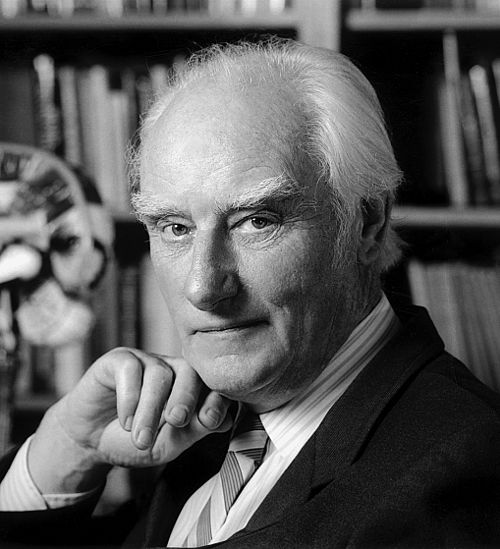
Francis Crick crop

Diagram that emphasises the phosphate backbone of DNA. Watson and Crick first made helical models with the phosphates at the centre of the helices.

Crick and Watson DNA model built in 1953 was reconstructed largely from its original pieces in 1973 and donated to the National Science Museum in London.

Results from an fMRI experiment in which people made a conscious decision about a visual stimulus. The small region of the brain coloured orange shows patterns of activity that correlate with the decision making process. Crick stressed the importance of finding new methods to probe human brain function.