Patch clamp
Videos
Page
The patch clamp technique is a laboratory technique in electrophysiology used to study ionic currents in individual isolated living cells, tissue sections, or patches of cell membrane. The technique is especially useful in the study of excitable cells such as neurons, cardiomyocytes, muscle fibers, and pancreatic beta cells, and can also be applied to the study of bacterial ion channels in specially prepared giant spheroplasts.
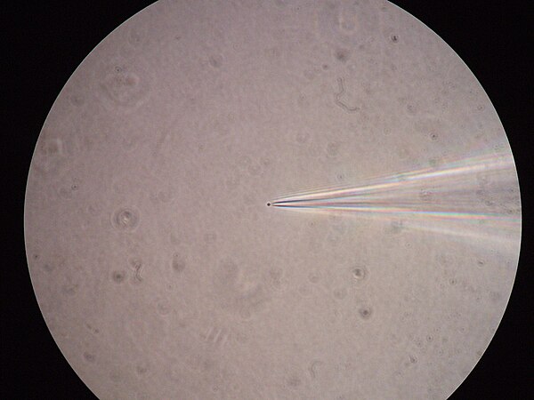
A bacterial spheroplast patched with a glass pipette

Patch clamp of a nerve cell within a slice of brain tissue. The pipette in the photograph has been marked with a slight blue color.
Electrophysiology
Videos
Page
Electrophysiology is the branch of physiology that studies the electrical properties of biological cells and tissues. It involves measurements of voltage changes or electric current or manipulations on a wide variety of scales from single ion channel proteins to whole organs like the heart. In neuroscience, it includes measurements of the electrical activity of neurons, and, in particular, action potential activity. Recordings of large-scale electric signals from the nervous system, such as electroencephalography, may also be referred to as electrophysiological recordings. They are useful for electrodiagnosis and monitoring.

A schematic diagram showing a field potential recording from rat hippocampus. At the left is a schematic diagram of a presynaptic terminal and postsynaptic neuron. This is meant to represent a large population of synapses and neurons. When the synapse releases glutamate onto the postsynaptic cell, it opens ionotropic glutamate receptor channels. The net flow of current is inward, so a current sink is generated. A nearby electrode (#2) detects this as a negativity. An