In histology, histopathology, and clinical pathology, Perls Prussian blue is a commonly used method to detect the presence of iron in tissue or cell samples. Perls Prussian Blue derives its name from the German pathologist Max Perls (1843–1881), who described the technique in 1867. The method does not involve the application of a dye, but rather causes the pigment Prussian blue to form directly within the tissue. The method stains mostly iron in the ferric state which includes ferritin and hemosiderin, rather than iron in the ferrous state.
Cerebrospinal fluid specimen stained with Perls Prussian blue showing iron containing macrophage (stained blue) surrounded by erythrocytes (stained red)
Section of liver stained with Perls Prussian blue, showing iron accumulations (blue) consistent with homozygous genetic hemochromatosis
Perls Prussian blue components: potassium ferrocyanide and hydrochloric acid.
Perls Prussian blue stained section of liver biopsy showing hemosiderosis
Histology,
also known as microscopic anatomy or microanatomy, is the branch of biology that studies the microscopic anatomy of biological tissues. Histology is the microscopic counterpart to gross anatomy, which looks at larger structures visible without a microscope. Although one may divide microscopic anatomy into organology, the study of organs, histology, the study of tissues, and cytology, the study of cells, modern usage places all of these topics under the field of histology. In medicine, histopathology is the branch of histology that includes the microscopic identification and study of diseased tissue. In the field of paleontology, the term paleohistology refers to the histology of fossil organisms.
Histologic specimen being placed on the stage of an optical microscope.
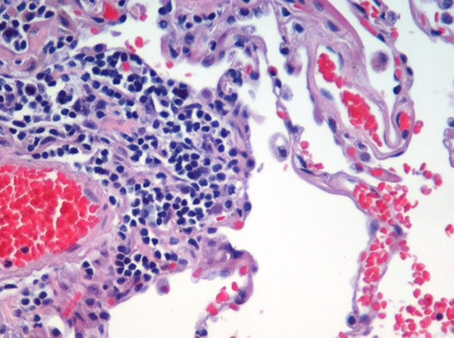
Human lung tissue stained with hematoxylin and eosin as seen under a microscope.
Histologic section of a plant stem (Alliaria petiolata).
Histologic section of a fossilized invertebrate. Ordovician bryozoan.