Radiographer
Videos
Radiographers are healthcare professionals who perform medical imaging and radiation therapy. Medical imaging is used for the diagnosis of pathology, while radiation therapy is used for treatment.
A radiographer with a radiographic examination table and X-Ray tube


Taking an X-ray image with early Crookes tube apparatus, late 1800s
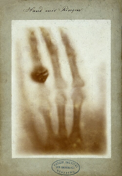
The first radiograph

A modern diagnostic radiology acquisition console. The evolution of allied health roles has expanded a radiographer's clinical domain to include advanced computed tomography (CT) slice profiling and multiplanar volume reconstruction.
Radiology
Videos
Radiology is the medical specialty that uses medical imaging to diagnose diseases and guide treatment within the bodies of humans and other animals. It began with radiography, but today it includes al
Radiographer
…between the roles that we now differentiate as radiologic technologist (a technician in an allied health professions who obtains the images) versus radiologist (a physician who interprets them). By the 1930s and 1940s, as it became increasingly apparent that proper interpretation of the images required not…

A radiologist interpreting magnetic resonance imaging

Image from a CT scan of the brain

A modern diagnostic imaging suite in Navi Mumbai, India, featuring a computed tomography (CT) scanner utilized for specialized cranial, abdominal, and angiographic medical evaluations.

MRI of the knee