Sarfus is an optical quantitative imaging technique based on the association of:an upright or inverted optical microscope in crossed polarization configuration and
specific supporting plates – called surfs – on which the sample to observe is deposited.
3D sarfus image of a DNA biochip.
Observation with standard optical microscope between cross polarizers of Langmuir-Blodgett layers (bilayer thickness: 5.4 nm) on silicon wafer and on surf
Light polarisation after reflexion on a surf (0) and on nanoscale sample on a surf (1).
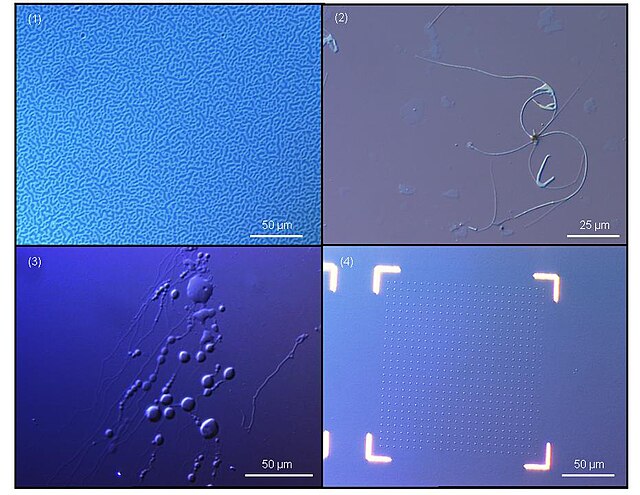
Sarfus images of nanostructures: 1. Copolymer film microstructuration (73 nm), 2. Carbon nanotube bundles, 3. Lipid vesicles in aqueous solutions, 4. Nanopatterning of gold dots (50 nm3).
The optical microscope, also referred to as a light microscope, is a type of microscope that commonly uses visible light and a system of lenses to generate magnified images of small objects. Optical microscopes are the oldest design of microscope and were possibly invented in their present compound form in the 17th century. Basic optical microscopes can be very simple, although many complex designs aim to improve resolution and sample contrast.
Scientist using an optical microscope in a laboratory
A miniature USB microscope
The oldest published image known to have been made with a microscope: bees by Francesco Stelluti, 1630
Basic optical transmission microscope elements (1990s)


