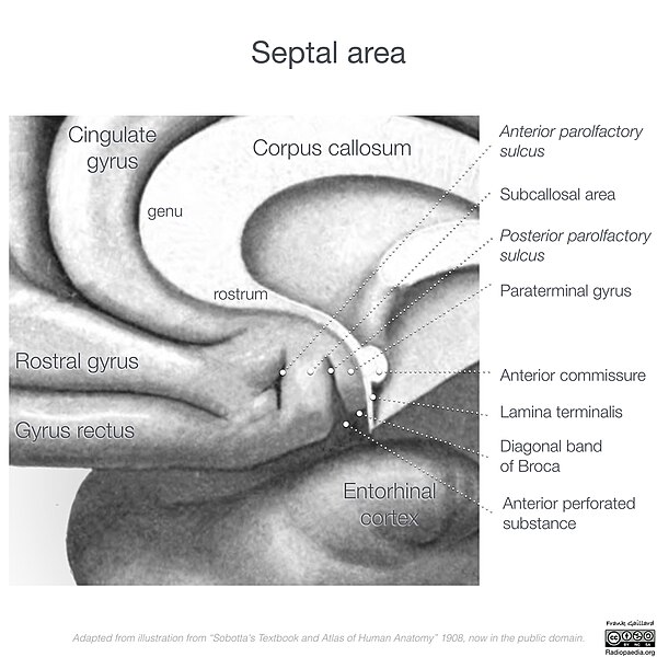
The septal area, consisting of the lateral septum and medial septum, is an area in the lower, posterior part of the medial surface of the frontal lobe, and refers to the nearby septum pellucidum.
Lateral and medial septal nuclei in the septal area of the mouse brain
Detail of drawing showing components of septal area below corpus callosum.
The nucleus accumbens is a region in the basal forebrain rostral to the preoptic area of the hypothalamus. The nucleus accumbens and the olfactory tubercle collectively form the ventral striatum. The ventral striatum and dorsal striatum collectively form the striatum, which is the main component of the basal ganglia. The dopaminergic neurons of the mesolimbic pathway project onto the GABAergic medium spiny neurons of the nucleus accumbens and olfactory tubercle. Each cerebral hemisphere has its own nucleus accumbens, which can be divided into two structures: the nucleus accumbens core and the nucleus accumbens shell. These substructures have different morphology and functions.
Nucleus accumbens of the mouse brain
Tuning of appetitive and defensive reactions in the nucleus accumbens shell. (Above) AMPA blockade requires D1 function in order to produce motivated behaviors, regardless of valence, and D2 function to produce defensive behaviors. GABA agonism, on the other hand, does not require dopamine receptor function.(Below)The expansion of the anatomical regions that produce defensive behaviors under stress, and appetitive behaviors in the home environment produced by AMPA antagonism. This flexibility is less evident with GABA agonism.
Sagittal MRI slice with highlighting (red) indicating the nucleus accumbens


