Spindle apparatus
Videos
In cell biology, the spindle apparatus is the cytoskeletal structure of eukaryotic cells that forms during cell division to separate sister chromatids between daughter cells. It is referred to as the

Micrograph showing condensed chromosomes in blue, kinetochores in pink, and microtubules in green during metaphase of mitosis
Mitosis
Videos
Mitosis is a part of the cell cycle in eukaryotic cells in which replicated chromosomes are separated into two new nuclei. Cell division by mitosis is an equational division which gives rise to geneti
Spindle apparatus
…eukaryotic cells that forms during cell division to separate sister chromatids between daughter cells. It is referred to as the mitotic spindle during mitosis, a process that produces genetically identical daughter cells, or the meiotic spindle during meiosis, a process that produces gametes with half the…

Interphase nucleus (left), condensing chromosomes (middle) and condensed chromosomes (right)

A cell in late metaphase. All chromosomes (blue) but one have arrived at the metaphase plate.
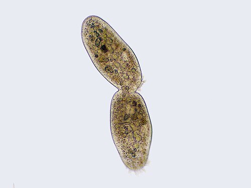
Ciliate undergoing cytokinesis, with the cleavage furrow being clearly visible

An abnormal (tripolar) mitosis (12 o'clock position) in a precancerous lesion of the stomach (H&E stain)