Spliceosome
Videos
A spliceosome is a large ribonucleoprotein complex found primarily within the nucleus of eukaryotic cells. The spliceosome is assembled from small nuclear RNAs and numerous proteins. Small nuclear RNA
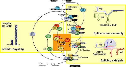
Spliceosomal splicing cycle

Figure 1. Above are electron microscopy fields of negatively stained yeast (Saccharomyces cerevisiae) tri-snRNPs. Below left is a schematic illustration of the interaction of tri-snRNP proteins with the U4/U6 snRNA duplex. Below right is a cartoon model of the yeast tri-snRNP with shaded areas corresponding to U5 (gray), U4/U6 (orange) and the linker region (yellow).
Minor spliceosome
Videos
The minor spliceosome is a ribonucleoprotein complex that catalyses the removal of an atypical class of spliceosomal introns from messenger RNAs in some clades of eukaryotes. This process is called no
Spliceosome
…structure is mutually exclusive with the 3' half of an internal 5' stem loop region of U2 snRNA. Some eukaryotes have a second spliceosome, the so-called minor spliceosome. A group of less abundant snRNAs, U11, U12, U4atac, and U6atac, together with U5, are subunits of the minor spliceosome that splices a rare class of…

A comparison between major and minor splicing mechanisms

U1 and U11 can be folded similarly