The Stramenopiles, also called Heterokonts, are a clade of organisms distinguished by the presence of stiff tripartite external hairs. In most species, the hairs are attached to flagella, in some they are attached to other areas of the cellular surface, and in some they have been secondarily lost. Stramenopiles represent one of the three major clades in the SAR supergroup, along with Alveolata and Rhizaria.
Two living C. roenbergensis. Light micrograph. The cells are about 6 µm long. The anterior flagellum beats with an undulating pattern, the posterior (recurrent or smooth) flagellum usually holds the cell to the substrate.
Giant kelp, Macrocystis pyrifera, an example of a multicellular stramenopile, is a large seaweed, up to 45 metres (150 feet) long, in the Phaeophyceae, within the Gyrista.
Electron micrograph of the protist Paraphysomonas butcheri. It illustrates the stramenopile property – of having stiff hairs. The hairs attach to one longer flagellum, the other is without hairs (an arrangement also called 'heterokont', meaning "unequal"). The body of the flagellate is coated with delicate scales. Paraphysomonas feeds on bacteria, two of which lie near the hairy flagellum.
A flagellum is a hairlike appendage that protrudes from certain plant and animal sperm cells, from fungal spores (zoospores), and from a wide range of microorganisms to provide motility. Many protists with flagella are known as flagellates.
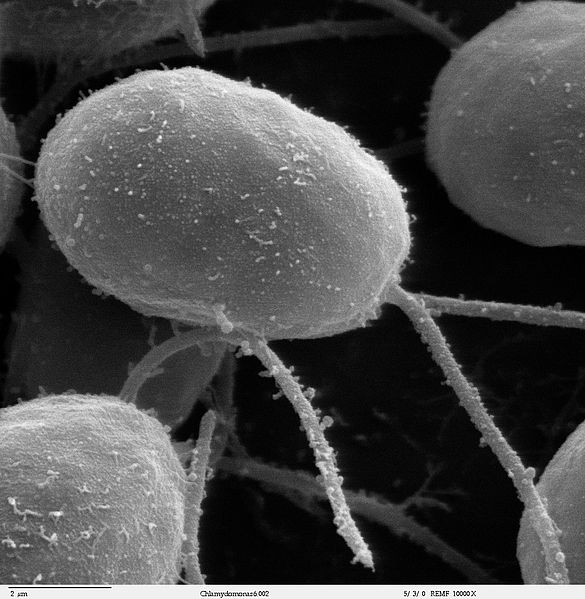
SEM image of flagellated eukaryote Chlamydomonas sp. (10000×)
Bacterial flagellar motor assembly: Shown here is the C-ring at the base with FliG in red, FliM in yellow, and FliN in shades of purple; the MS-ring in blue; the MotAB in brown; the LP-ring in pink; and the rod in gray.
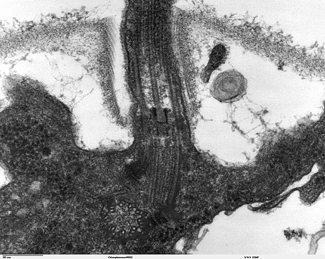
Longitudinal section through the flagella area in Chlamydomonas reinhardtii. In the cell apex is the basal body that is the anchoring site for a flagellum. Basal bodies originate from and have a substructure similar to that of centrioles, with nine peripheral microtubule triplets (see structure at bottom center of image).
The "9+2" structure is visible in this cross-section micrograph of an axoneme.