Thorax
Videos
Page
The thorax or chest is a part of the anatomy of mammals and other tetrapod animals located between the neck and the abdomen. In insects, crustaceans, and the extinct trilobites, the thorax is one of the three main divisions of the creature's body, each of which is in turn composed of multiple segments.

X-ray image of the human chest showing the internal anatomy of the rib cage, lungs and heart as well as the inferior thoracic border–made up of the diaphragm.
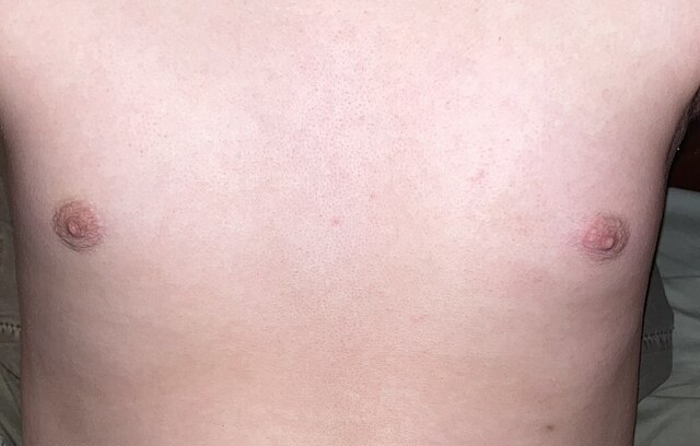
Adolescent male chest and nipples.
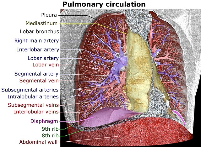
Volume rendering of a high resolution computed tomography of the thorax. The anterior thoracic wall, the airways and the pulmonary vessels anterior to the root of the lung have been digitally removed in order to visualize the different levels of the pulmonary circulation.
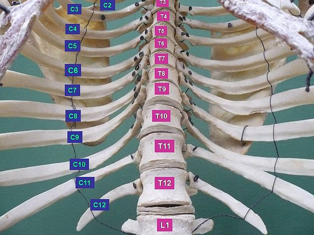
Thorax. Anterior view.
Anatomy
Videos
Page
Anatomy is the branch of morphology concerned with the study of the internal structure of organisms and their parts. Anatomy is a branch of natural science that deals with the structural organization of living things. It is an old science, having its beginnings in prehistoric times. Anatomy is inherently tied to developmental biology, embryology, comparative anatomy, evolutionary biology, and phylogeny, as these are the processes by which anatomy is generated, both over immediate and long-term timescales. Anatomy and physiology, which study the structure and function of organisms and their parts respectively, make a natural pair of related disciplines, and are often studied together. Human anatomy is one of the essential basic sciences that are applied in medicine, and is often studied alongside physiology.
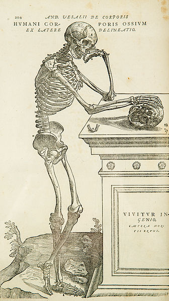
One of the large, detailed illustrations in Andreas Vesalius's De humani corporis fabrica 16th century, marking the rebirth of anatomy
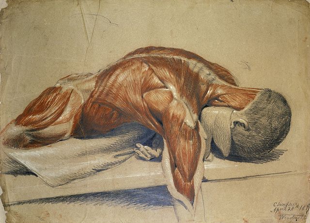
A dissected body, lying prone on a table, by Charles Landseer

Hyaline cartilage at high magnification (H&E stain)

Gastric mucosa at low magnification (H&E stain)