White matter refers to areas of the central nervous system (CNS) that are mainly made up of myelinated axons, also called tracts. Long thought to be passive tissue, white matter affects learning and brain functions, modulating the distribution of action potentials, acting as a relay and coordinating communication between different brain regions.
Micrograph showing white matter with its characteristic fine meshwork-like appearance (left of image – lighter shade of pink) and grey matter, with the characteristic neuronal cell bodies (right of image – dark shade of pink). HPS stain.
Human brain right dissected lateral view, showing grey matter (the darker outer parts), and white matter (the inner and prominently whiter parts).
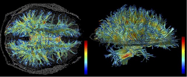
White matter structure of human brain (taken by MRI).
Myelin is a lipid-rich material that surrounds nerve cell axons to insulate them and increase the rate at which electrical impulses pass along the axon. The myelinated axon can be likened to an electrical wire with insulating material (myelin) around it. However, unlike the plastic covering on an electrical wire, myelin does not form a single long sheath over the entire length of the axon. Rather, myelin ensheaths the axon segmentally: in general, each axon is encased in multiple long sheaths with short gaps between, called nodes of Ranvier. At the nodes of Ranvier, which are approximately one thousandth of a mm in length, the axon's membrane is bare of myelin.
Transmission electron micrograph of a cross-section of a myelinated PNS axon, generated at the Electron Microscopy Facility at Trinity College, Hartford, Connecticut
Myelin formed by Schwann cells in the PNS