Pathology
Videos
Page
Pathology is the study of disease and injury. The word pathology also refers to the study of disease in general, incorporating a wide range of biology research fields and medical practices. However, when used in the context of modern medical treatment, the term is often used in a narrower fashion to refer to processes and tests that fall within the contemporary medical field of "general pathology", an area that includes a number of distinct but inter-related medical specialties that diagnose disease, mostly through analysis of tissue and human cell samples. Idiomatically, "a pathology" may also refer to the predicted or actual progression of particular diseases, and the affix pathy is sometimes used to indicate a state of disease in cases of both physical ailment and psychological conditions. A physician practicing pathology is called a pathologist.
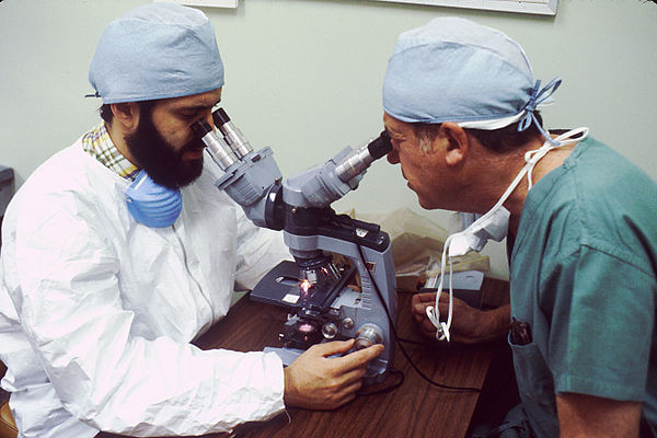
A pathologist examines a tissue section for evidence of cancerous cells while a surgeon observes.

The advent of the microscope was one of the major developments in the history of pathology. Here researchers at the Centers for Disease Control in 1978 examine cultures containing Legionella pneumophila, the pathogen responsible for Legionnaire's disease.
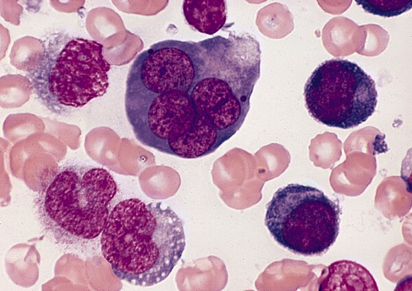
A bone marrow smear from a case of erythroleukemia. The large cell in the top center is an abnormal erythroblast: it is multinucleated, with megaloblastoid nuclear chromatin. This is diagnostic of erythroleukemia.

A malignant melanoma can often be suspected from sight, but confirmation of the diagnosis or outright removal requires a biopsy.
Anatomical pathology
Videos
Page
Anatomical pathology (Commonwealth) or anatomic pathology (U.S.) is a medical specialty that is concerned with the diagnosis of disease based on the macroscopic, microscopic, biochemical, immunologic and molecular examination of organs and tissues. Over the 20th century, surgical pathology has evolved tremendously: from historical examination of whole bodies (autopsy) to a more modernized practice, centered on the diagnosis and prognosis of cancer to guide treatment decision-making in oncology. Its modern founder was the Italian scientist Giovanni Battista Morgagni from Forlì.
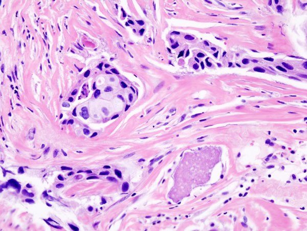
Histopathology: microscopic appearance of invasive ductal carcinoma of the breast. The slide is stained with Haematoxylin & Eosin.

Histopathology: microscopic appearance of invasive ductal carcinoma of the breast. The slide is stained with an antibody (immunohistochemistry) against the oncogene Her2neu. The dark-brown reaction indicates that this tumor over-expresses this gene.
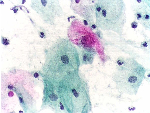
Cytopathology: microscopic appearance of a Pap test. The pink cell at the center with a large nucleus is abnormal, compatible with low-grade dysplasia.

Autopsy: a brain surrounded by pus (the yellow-greyish coat around the brain, under the dura lifted by the forceps), the result of bacterial meningitis.