Visual cortex
Videos
Page
The visual cortex of the brain is the area of the cerebral cortex that processes visual information. It is located in the occipital lobe. Sensory input originating from the eyes travels through the lateral geniculate nucleus in the thalamus and then reaches the visual cortex. The area of the visual cortex that receives the sensory input from the lateral geniculate nucleus is the primary visual cortex, also known as visual area 1 (V1), Brodmann area 17, or the striate cortex. The extrastriate areas consist of visual areas 2, 3, 4, and 5.

Micrograph showing the visual cortex (pink). The pia mater and arachnoid mater including blood vessels are seen at the top of the image. Subcortical white matter (blue) is seen at the bottom of the image. HE-LFB stain.
Cerebral cortex
Videos
Page
The cerebral cortex, also known as the cerebral mantle, is the outer layer of neural tissue of the cerebrum of the brain in humans and other mammals. It is the largest site of neural integration in the central nervous system, and plays a key role in attention, perception, awareness, thought, memory, language, and consciousness. The cerebral cortex is the part of the brain responsible for cognition.
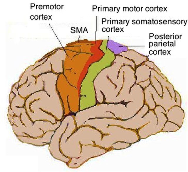
Lateral view of cerebrum showing several cortices
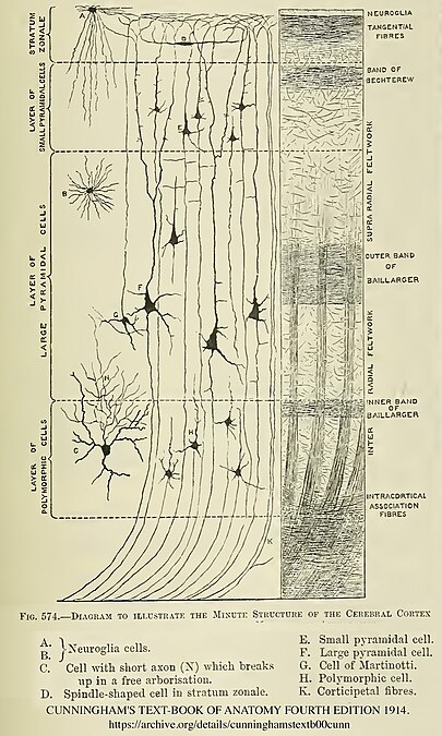
Diagram of layers pattern. Cells grouped on left, axonal layers on right.
Micrograph showing the visual cortex (predominantly pink). Subcortical white matter (predominantly blue) is seen at the bottom of the image. HE-LFB stain.

Cortical blood supply