Adaptive immune system
Videos
The adaptive immune system, also known as the acquired immune system, or specific immune system is a subsystem of the immune system that is composed of specialized, systemic cells and processes that eliminate pathogens or prevent their growth. The acquired immune system is one of the two main immunity strategies found in vertebrates.
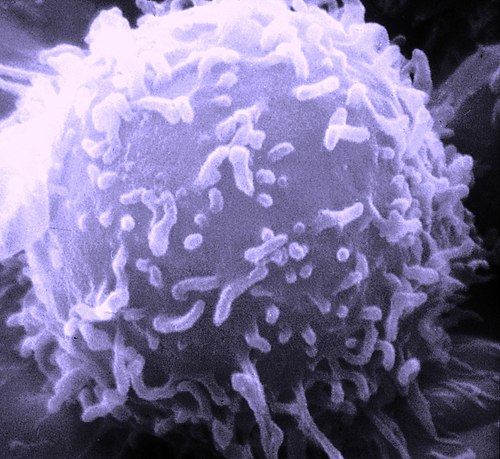
A scanning electron microscope image of a single human lymphocyte
Lymphocyte
Videos
A lymphocyte is a type of white blood cell (leukocyte) in the immune system of most vertebrates. Lymphocytes include T cells, B cells, and innate lymphoid cells, of which natural killer cells are an important subtype. They are the main type of cell found in lymph, which prompted the name "lymphocyte". Lymphocytes make up between 18% and 42% of circulating white blood cells.

Scanning electron micrograph of a human T cell

A stained lymphocyte surrounded by red blood cells viewed using a light microscope
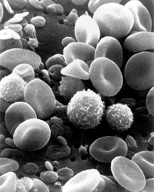
A scanning electron microscope image of normal circulating human blood showing red blood cells, several types of white blood cells including lymphocytes, a monocyte, a neutrophil and many small disc-shaped platelets

Several lymphocytes seen collected around a tuberculous granuloma