Alpha helix
Videos
An alpha helix is a sequence of amino acids in a protein that are twisted into a coil.

Contrast of helix end views between α (offset squarish) vs 310 (triangular)
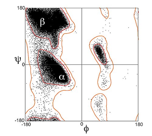
Ramachandran plot (φ, ψ plot), with data points for α-helical residues forming a dense diagonal cluster below and left of center, around the global energy minimum for backbone conformation.

An α-helix in ultrahigh-resolution electron density contours, with oxygen atoms in red, nitrogen atoms in blue, and hydrogen bonds as green dotted lines (PDB file 2NRL, 17–32). The N-terminus is at the top, here.

Leucine zipper coiled-coil helices & DNA-binding helices: transcription factor Max (PDB file 1HLO)
Helix
Videos
A helix is a shape like a cylindrical coil spring or the thread of a machine screw. It is a type of smooth skew curve with tangent lines at a constant angle to a fixed axis. Helices are important in b
Alpha helix
…An alpha helix (or α-helix) is a sequence of amino acids in a protein that are twisted into a coil (a helix). The alpha helix is the most common structural arrangement in the secondary structure of proteins. It is also the most extreme type of local structure…

(l-r) Tension, compression and torsion coil springs

A machine screw

Crystal structure of a folded molecular helix reported by Lehn et al.
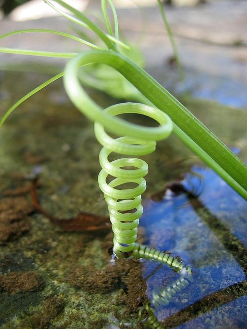
A natural left-handed helix, made by a climber plant