Astrocyte
Videos
Astrocytes, also known collectively as astroglia, are characteristic star-shaped glial cells in the brain and spinal cord. They perform many functions, including biochemical control of endothelial cells that form the blood–brain barrier, provision of nutrients to the nervous tissue, maintenance of extracellular ion balance, regulation of cerebral blood flow, and a role in the repair and scarring process of the brain and spinal cord following infection and traumatic injuries. The proportion of astrocytes in the brain is not well defined; depending on the counting technique used, studies have found that the astrocyte proportion varies by region and ranges from 20% to around 40% of all glia. Another study reports that astrocytes are the most numerous cell type in the brain. Astrocytes are the major source of cholesterol in the central nervous system. Apolipoprotein E transports cholesterol from astrocytes to neurons and other glial cells, regulating cell signaling in the brain. Astrocytes in humans are more than twenty times larger than in rodent brains, and make contact with more than ten times the number of synapses.

An astrocyte from a rat brain grown in tissue culture and stained with antibodies to GFAP (red) and vimentin (green). Both proteins are present in large amounts in the intermediate filaments of this cell, so the cell appears yellow. The blue material shows DNA visualized with DAPI stain, and reveals the nucleus of the astrocyte and of other cells. Image courtesy of EnCor Biotechnology Inc.
Astrocytes (green) in the context of neurons (red) in a mouse cortex cell culture

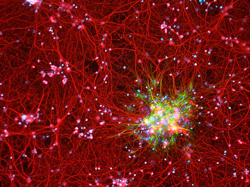
Astrocytes are depicted in red. Cell nuclei are depicted in blue. Astrocytes were obtained from brains of newborn mice.
Vimentin
Videos
Vimentin is a structural protein that in humans is encoded by the VIM gene. Its name comes from the Latin vimentum which refers to an array of flexible rods.

Immunofluorescence staining pattern of vimentin antibodies. Produced by incubating vimentin primary antibodies and FITC labelled secondary antibodies with HEp-20-10 cells.

Immunofluorescence staining of HeLa Cells with antibody to reveal vimentin containing intermediate filaments in green and antibody to LAMP1 to reveal lysosomes in red. Nuclear DNA is seen in blue. Antibodies and image courtesy EnCor Biotechnology Inc.