In molecular biology, biochips are engineered substrates that can host large numbers of simultaneous biochemical reactions. One of the goals of biochip technology is to efficiently screen large numbers of biological analytes, with potential applications ranging from disease diagnosis to detection of bioterrorism agents. For example, digital microfluidic biochips are under investigation for applications in biomedical fields. In a digital microfluidic biochip, a group of (adjacent) cells in the microfluidic array can be configured to work as storage, functional operations, as well as for transporting fluid droplets dynamically.
Hundreds of gel drops are visible on the biochip.
Figure 1. Biochips are a platform that require, in addition to microarray technology, transduction and signal processing technologies to output the results of sensing experiments.
3D Sarfus image of a DNA biochip
Sarfus is an optical quantitative imaging technique based on the association of:an upright or inverted optical microscope in crossed polarization configuration and
specific supporting plates – called surfs – on which the sample to observe is deposited.
3D sarfus image of a DNA biochip.
Observation with standard optical microscope between cross polarizers of Langmuir-Blodgett layers (bilayer thickness: 5.4 nm) on silicon wafer and on surf
Light polarisation after reflexion on a surf (0) and on nanoscale sample on a surf (1).
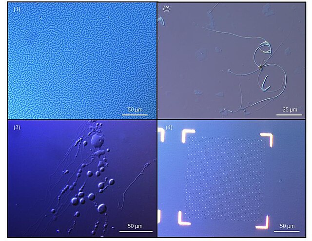
Sarfus images of nanostructures: 1. Copolymer film microstructuration (73 nm), 2. Carbon nanotube bundles, 3. Lipid vesicles in aqueous solutions, 4. Nanopatterning of gold dots (50 nm3).


