Capillary
Videos
A capillary is a small blood vessel, from 5 to 10 micrometres in diameter, and is part of the microcirculation system. Capillaries are microvessels and the smallest blood vessels in the body. They are composed of only the tunica intima, consisting of a thin wall of simple squamous endothelial cells. They are the site of the exchange of many substances from the surrounding interstitial fluid, and they convey blood from the smallest branches of the arteries (arterioles) to those of the veins (venules). Other substances which cross capillaries include water, oxygen, carbon dioxide, urea, glucose, uric acid, lactic acid and creatinine. Lymph capillaries connect with larger lymph vessels to drain lymphatic fluid collected in microcirculation.
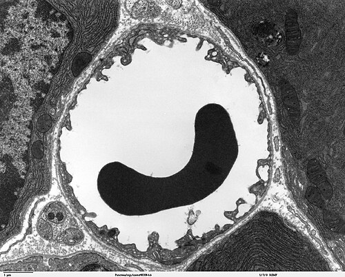
Transmission electron microscope image of a cross-section of a capillary occupied by a red blood cell

A simplified illustration of a capillary network

Types of capillaries: (left) continuous with no big gaps, (center) fenestrated with small pores, and (right) sinusoidal (or 'discontinuous') with intercellular gaps

Scanning electron micrograph of a liver sinusoid with fenestrated endothelial cells. Fenestrae are approximately 100 nm in diameter.
Extracellular fluid
Videos
In cell biology, extracellular fluid (ECF) denotes all body fluid outside the cells of any multicellular organism. Total body water in healthy adults is about 50–60% of total body weight; women and the obese typically have a lower percentage than lean men. Extracellular fluid makes up about one-third of body fluid, the remaining two-thirds is intracellular fluid within cells. The main component of the extracellular fluid is the interstitial fluid that surrounds cells.

The distribution of the total body water in mammals between the intracellular compartment and the extracellular compartment, which is, in turn, subdivided into interstitial fluid and smaller components, such as the blood plasma, the cerebrospinal fluid and lymph