Cardiac cycle
Videos
The cardiac cycle is the performance of the human heart from the beginning of one heartbeat to the beginning of the next. It consists of two periods: one during which the heart muscle relaxes and refi
The cycle diagram depicts one heartbeat of the continuously repeating cardiac cycle, namely: ventricular diastole followed by ventricular systole, etc.—while coordinating with atrial systole followed by atrial diastole, etc. The cycle also correlates to key electrocardiogram tracings: the T wave (which indicates ventricular diastole); the P wave (atrial systole); and the QRS 'spikes' complex (ventricular systole)—all shown as color purple-in-black segments.

Mitral valve
Videos
The mitral valve, also known as the bicuspid valve or left atrioventricular valve, is one of the four heart valves. It has two cusps or flaps and lies between the left atrium and the left ventricle of

Operative view of the mitral valve with a chordal rupture "fail" of the anterior leaflet
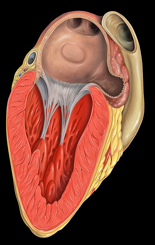
The left side of the heart. The mitral valve, as well as the chordae tendinae are visible as white strings. These connect to the papillary muscles visible attaching to the muscular ventricle.

Mitral valve, viewed in a cadaver specimen from within the left atrium.