Mitral valve
Videos
The mitral valve, also known as the bicuspid valve or left atrioventricular valve, is one of the four heart valves. It has two cusps or flaps and lies between the left atrium and the left ventricle of

Operative view of the mitral valve with a chordal rupture "fail" of the anterior leaflet
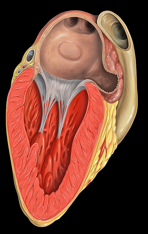
The left side of the heart. The mitral valve, as well as the chordae tendinae are visible as white strings. These connect to the papillary muscles visible attaching to the muscular ventricle.

Mitral valve, viewed in a cadaver specimen from within the left atrium.
Mitre
Videos
The mitre or miter is a type of headgear now known as the traditional, ceremonial headdress of bishops and certain abbots in traditional Christianity. Mitres are worn in the Catholic Church, Malankara
Mitral valve
…an ultrasound scan, which can reveal the size, anatomy and flow of blood through the valve. The word mitral comes from Latin, meaning "shaped like a mitre" (bishop's hat). The word bicuspid uses combining forms of bi-, from Latin, meaning "double", and cusp, meaning "point", reflecting the dual-flap shape…

A Catholic mitre from the 19th century, displayed in the Museo dell'Opera del Duomo in Pisa, Italy

The mitre of the Serbian Orthodox Bishop Georgije Đokić

The archbishop of the Church of Sweden, Martin Modéus, wearing a mitre as part of his liturgical vestment.

Mitre simplex traditional style: White damask with its white lappets ending in red fringes.