Connexin
Videos
Connexins, or gap junction proteins, are structurally related transmembrane proteins that assemble to form vertebrate gap junctions. An entirely different family of proteins, the innexins, forms gap j

Connexin-26 dodecamer. A gap junction, composed of twelve identical connexin proteins, six in the membrane of each cell. Each of these six units is a single polypeptide which passes the membrane four times (referred to as four-pass transmembrane proteins).

Life cycle and protein associations of connexins. Connexins are synthesized on ER-bound ribosomes and inserted into the ER cotranslationally. This is followed by oligomerization between the ER and trans-Golgi network (depending on the connexin type) into connexons, which are then delivered to the membrane via the actin or microtubule networks. Connexons may also be delivered to the plasma membrane by direct transfer from the rough ER. Upon insertion into the membrane, connexons may remain as hemichannels or they dock with compatible connexons on adjacent cells to form gap junctions. Newly delivered connexons are added to the periphery of pre-formed gap junctions, while the central "older" gap junction fragment are degraded by internalization of a double-membrane structure called an annular junction into one of the two cells, where subsequent lysosomal or proteasomal degradation occurs, or in some cases the connexons are recycled to the membrane (indicated by dashed arrow). During their life cycle, connexins associate with different proteins, including (1) cytoskeletal components as microtubules, actin, and actin-binding proteins α-
Astrocyte
Videos
Astrocytes, also known collectively as astroglia, are characteristic star-shaped glial cells in the brain and spinal cord. They perform many functions, including biochemical control of endothelial cel
Connexin
…atherosclerosis in mice. May have pathological effects. Also expressed between granulosa cells, which is required for proliferation. Normally expressed in astrocytes, also detected in most of the human astrocytomas and in the astroglial component of glioneuronal tumors. It is also the main cardiac connexin, found…
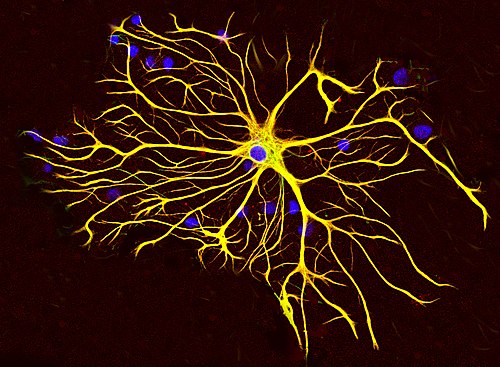
An astrocyte from a rat brain grown in tissue culture and stained with antibodies to GFAP (red) and vimentin (green). Both proteins are present in large amounts in the intermediate filaments of this cell, so the cell appears yellow. The blue material shows DNA visualized with DAPI stain, and reveals the nucleus of the astrocyte and of other cells. Image courtesy of EnCor Biotechnology Inc.
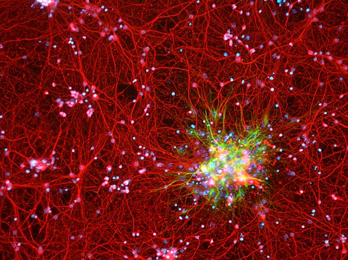
Astrocytes (green) in the context of neurons (red) in a mouse cortex cell culture

Astrocytes are depicted in red. Cell nuclei are depicted in blue. Astrocytes were obtained from brains of newborn mice.