The dentate nucleus is a cluster of neurons, or nerve cells, in the central nervous system that has a dentate – tooth-like or serrated – edge. It is located within the deep white matter of each cerebellar hemisphere, and it is the largest single structure linking the cerebellum to the rest of the brain. It is the largest and most lateral, or farthest from the midline, of the four pairs of deep cerebellar nuclei, the others being the globose and emboliform nuclei, which together are referred to as the interposed nucleus, and the fastigial nucleus. The dentate nucleus is responsible for the planning, initiation and control of voluntary movements. The dorsal region of the dentate nucleus contains output channels involved in motor function, which is the movement of skeletal muscle, while the ventral region contains output channels involved in nonmotor function, such as conscious thought and visuospatial function.
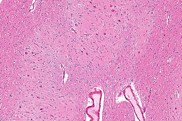
Micrograph of the dentate nucleus (pale pink). H&E stain.
White matter refers to areas of the central nervous system (CNS) that are mainly made up of myelinated axons, also called tracts. Long thought to be passive tissue, white matter affects learning and brain functions, modulating the distribution of action potentials, acting as a relay and coordinating communication between different brain regions.
Micrograph showing white matter with its characteristic fine meshwork-like appearance (left of image – lighter shade of pink) and grey matter, with the characteristic neuronal cell bodies (right of image – dark shade of pink). HPS stain.
Human brain right dissected lateral view, showing grey matter (the darker outer parts), and white matter (the inner and prominently whiter parts).
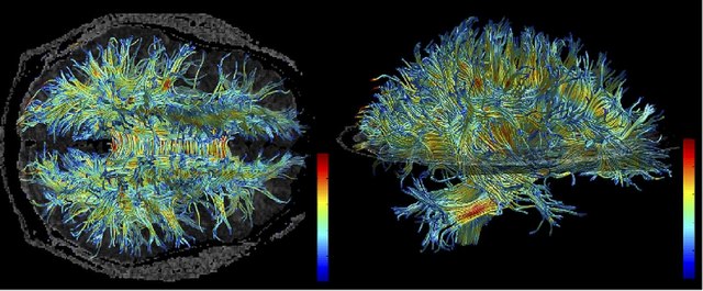
White matter structure of human brain (taken by MRI).