Electron diffraction
Videos
Photos
Electron diffraction is a generic term for phenomena associated with changes in the direction of electron beams due to elastic interactions with atoms. It occurs due to elastic scattering, when there is no change in the energy of the electrons. The negatively charged electrons are scattered due to Coulomb forces when they interact with both the positively charged atomic core and the negatively charged electrons around the atoms. The resulting map of the directions of the electrons far from the sample is called a diffraction pattern, see for instance Figure 1. Beyond patterns showing the directions of electrons, electron diffraction also plays a major role in the contrast of images in electron microscopes.
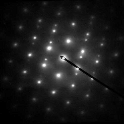
Figure 1: Selected area diffraction pattern of a twinned austenite crystal in a piece of steel

Figure 5: Replica built in 1980 by Ernst Ruska of the original electron microscope, in the Deutsches Museum in Munich

Figure 24: Gas electron diffraction pattern of benzene.

Image: Katódsugarak mágneses mezőben(1)
Cathode ray
Videos
Photos
Cathode rays or electron beams (e-beam) are streams of electrons observed in discharge tubes. If an evacuated glass tube is equipped with two electrodes and a voltage is applied, glass behind the positive electrode is observed to glow, due to electrons emitted from the cathode. They were first observed in 1859 by German physicist Julius Plücker and Johann Wilhelm Hittorf, and were named in 1876 by Eugen Goldstein Kathodenstrahlen, or cathode rays. In 1897, British physicist J. J. Thomson showed that cathode rays were composed of a previously unknown negatively charged particle, which was later named the electron. Cathode-ray tubes (CRTs) use a focused beam of electrons deflected by electric or magnetic fields to render an image on a screen.

A beam of cathode rays in a vacuum tube bent into a circle by a magnetic field generated by a Helmholtz coil. Cathode rays are normally invisible; in this demonstration Teltron tube, enough gas has been left in the tube for the gas atoms to luminesce when struck by the fast-moving electrons.
Crookes tube. The cathode (negative terminal) is on the right. The anode (positive terminal) is in the base of the tube at bottom.

Cathode rays travel from the cathode at the rear of the tube, striking the glass front, making it glow green by fluorescence. A metal cross in the tube casts a shadow, demonstrating that the rays travel in straight lines.

A magnet creates a horizontal magnetic field through the neck of the tube, bending the rays up, so the shadow of the cross is higher.