Endochondral ossification
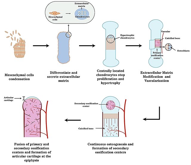
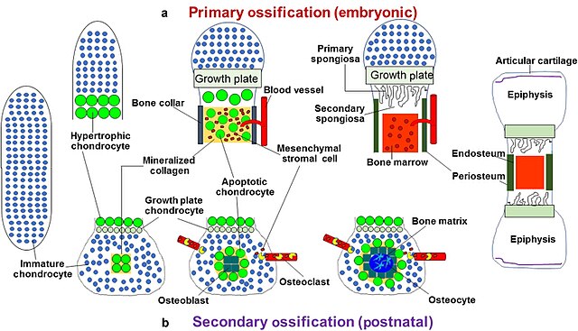
Endochondral ossification is one of the two essential pathways by which bone tissue is produced during fetal development of the mammalian skeletal system, the other pathway being intramembranous ossification. Both endochondral and intramembranous processes initiate from a precursor mesenchymal tissue, but their transformations into bone are different. In intramembranous ossification, mesenchymal tissue is directly converted into bone. On the other hand, endochondral ossification starts with mesenchymal tissue turning into an intermediate cartilage stage, which is eventually substituted by bone.
A schematic representation of endochondral ossification.
A schematic for long bone endochondral ossification.
Light micrograph of undecalcified epiphyseal plate showing endochondral ossification: healthy chondrocytes (top) become degenerating ones (bottom), characteristically displaying a calcified extracellular matrix.
Zones of endochondral ossification.
Intramembranous ossification
Intramembranous ossification is one of the two essential processes during fetal development of the gnathostome skeletal system by which rudimentary bone tissue is created.
Intramembranous ossification is also an essential process during the natural healing of bone fractures and the rudimentary formation of bones of the head.
Transmission electron micrograph of a mesenchymal stem cell that is displaying typical ultrastructural characteristics.
Light micrograph of a nidus consisting of osteoprogenitor cells that are displaying a prominent Golgi apparatus.
Light micrograph of a nidus consisting of osteoblasts, many are displaying a prominent Golgi apparatus, that have created osteoid at its center.
Light micrograph of an undecalcified nidus consisting of rudimentary bone tissue that is lined by numerous osteoblasts.


